Copyright
©The Author(s) 2023.
World J Gastrointest Surg. Sep 27, 2023; 15(9): 1901-1909
Published online Sep 27, 2023. doi: 10.4240/wjgs.v15.i9.1901
Published online Sep 27, 2023. doi: 10.4240/wjgs.v15.i9.1901
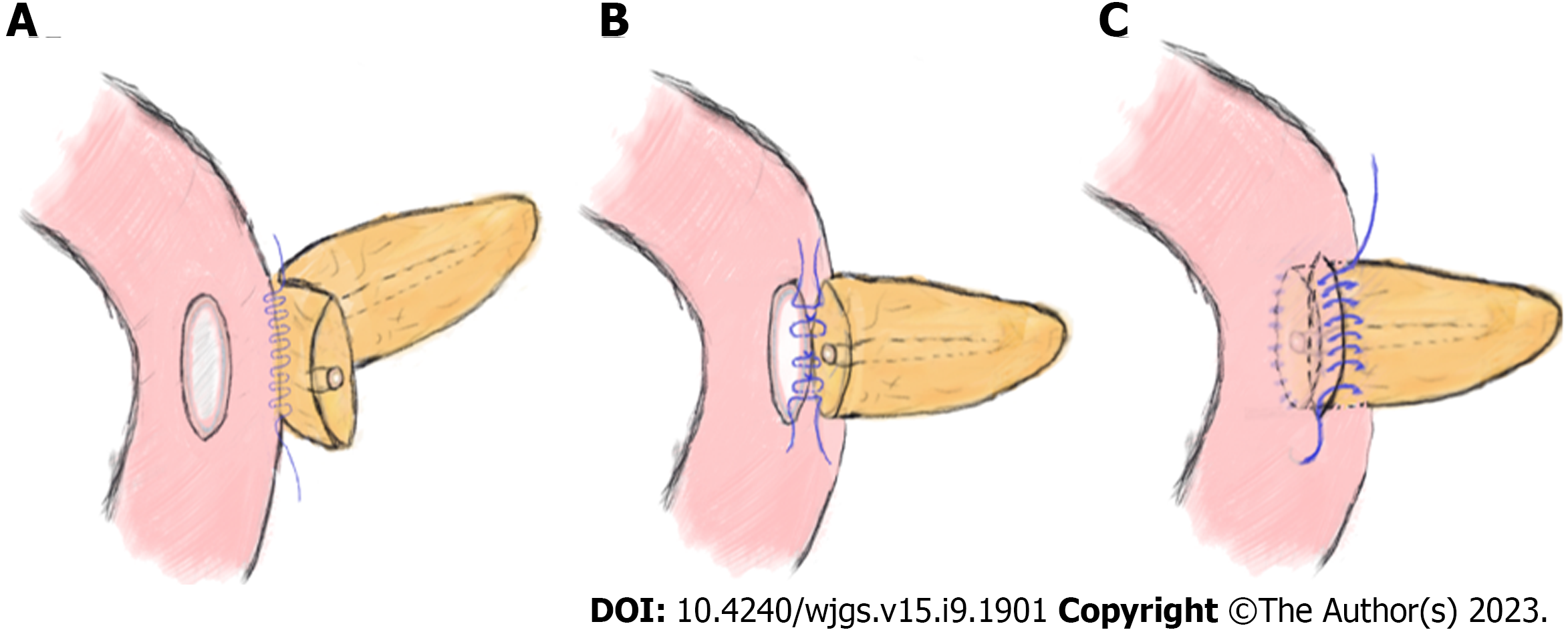
Figure 1 Schematic diagram of end-to-side invagination pancreaticojejunostomy.
A: Continuous suturing was performed between the rear side of the pancreatic stump (approximately 1.5 cm from its edge) and the jejunal seromuscular layer with a 3-0 Prolene slip line; B: Suture of the pancreatic margin and seromuscular layer of the jejunum intermittently; C: The pancreatic stump was inserted into the jejunum, and the anterior side of the pancreas and jejunal seromuscular layer were continuously sutured.
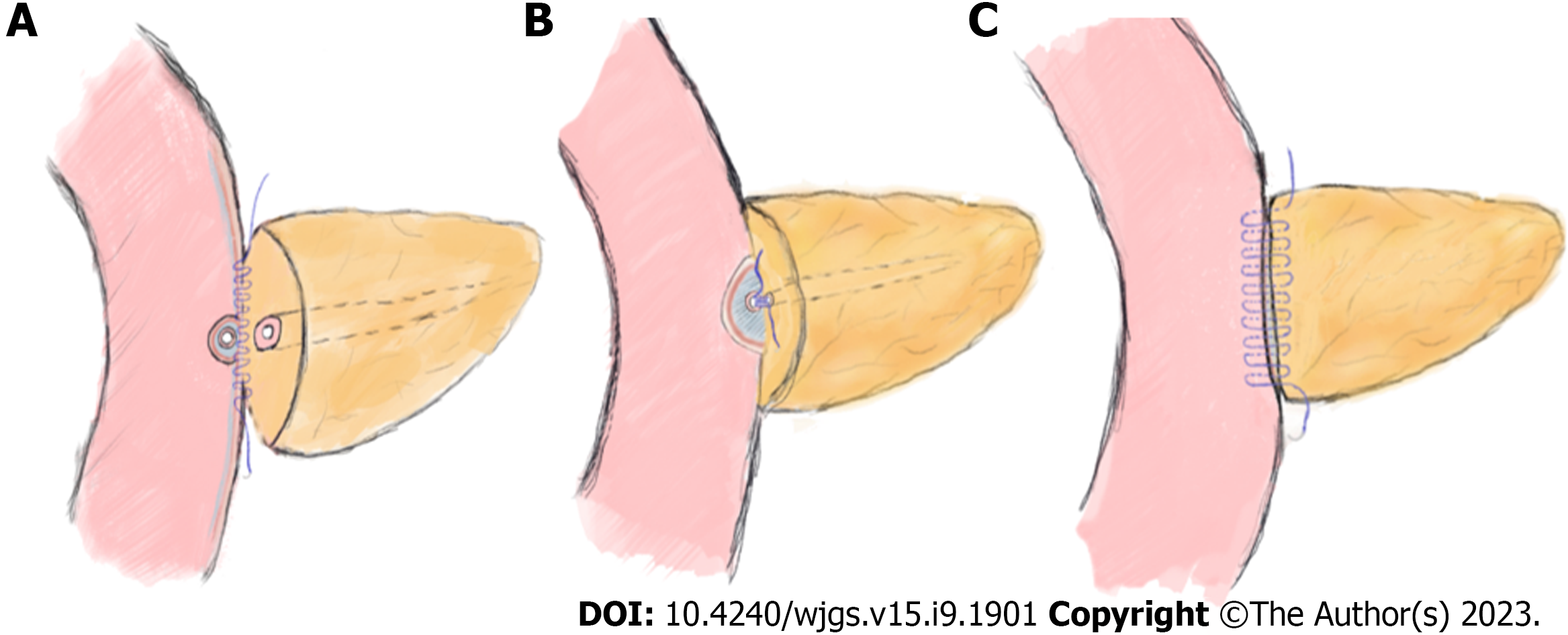
Figure 2 Schematic diagram of modified duct-to-mucosa pancreaticojejunostomy.
A: Perform continuous suturing between the rear edge of the pancreatic stump and jejunal seromuscular layer with a 3-0 Prolene suture; B: Sew 3-4 stiches continuously in the posterior wall of the pancreatic duct and the jejunal mucosa with 4-0 Prolene sutures; C: Continuous suturing was performed between the front edge of the pancreatic stump and the whole layer of the jejunum.
- Citation: Sun Y, Yu XF, Yao H, Xu S, Ma YQ, Chai C. Safety and feasibility of modified duct-to-mucosa pancreaticojejunostomy during pancreatoduodenectomy: A retrospective cohort study. World J Gastrointest Surg 2023; 15(9): 1901-1909
- URL: https://www.wjgnet.com/1948-9366/full/v15/i9/1901.htm
- DOI: https://dx.doi.org/10.4240/wjgs.v15.i9.1901


