Copyright
©The Author(s) 2023.
World J Gastrointest Surg. Aug 27, 2023; 15(8): 1831-1837
Published online Aug 27, 2023. doi: 10.4240/wjgs.v15.i8.1831
Published online Aug 27, 2023. doi: 10.4240/wjgs.v15.i8.1831
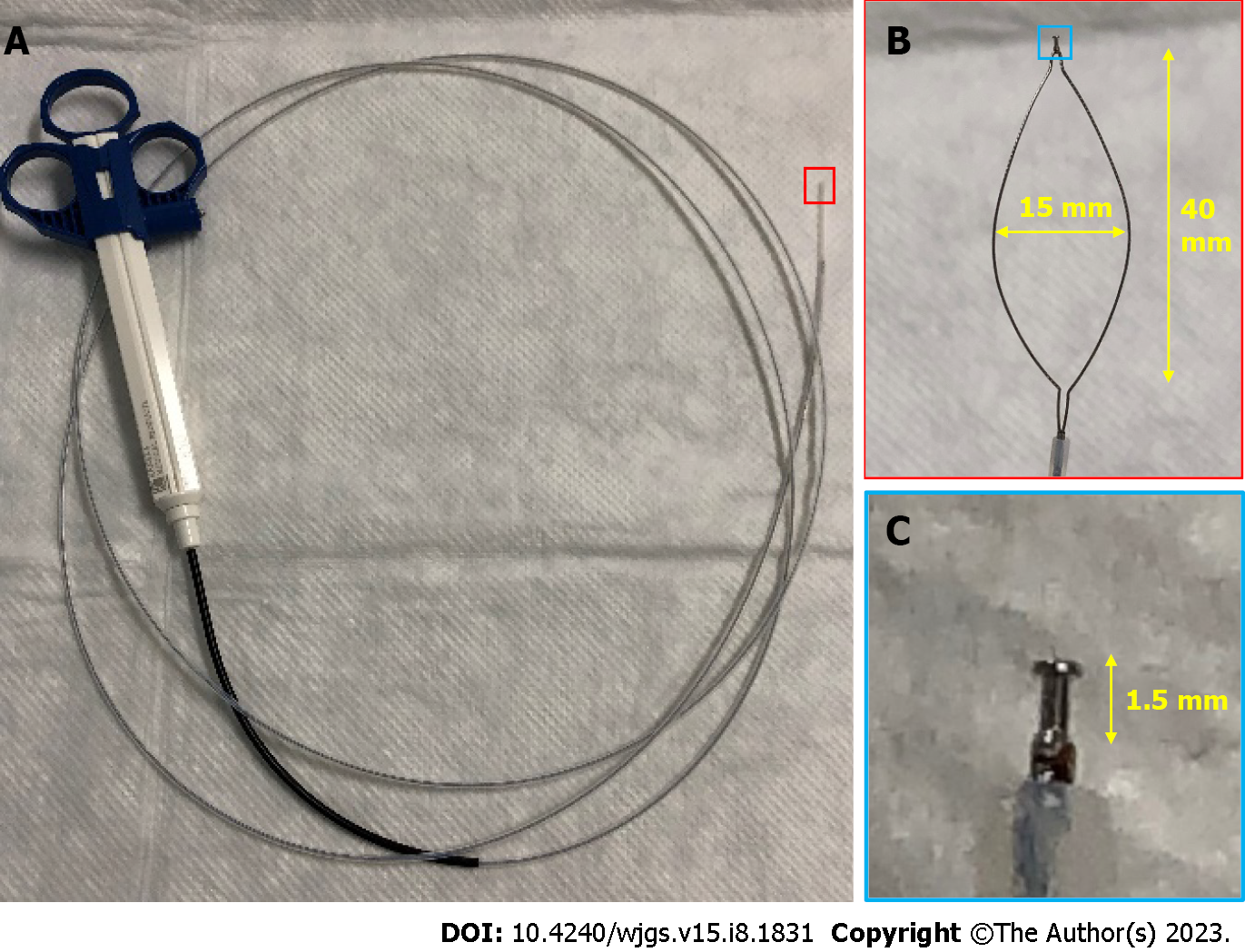
Figure 1 Details of the SOUTEN device, a multifunctional snare.
A: Full view of the SOUTEN device; B: The long diameter of the snare is 40 mm, and the short diameter of the snare is 15 mm; C: A 1.5-mm needle knife with a knob-shaped tip is attached to the top of the snare.
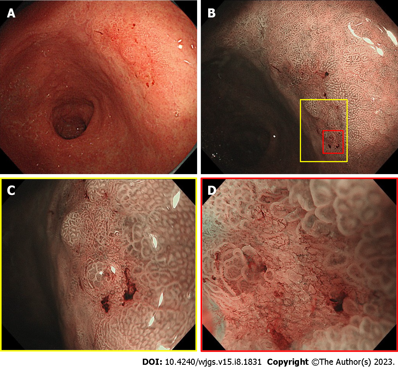
Figure 2 Endoscopic view of the 10-mm flat-depressed lesion on the posterior wall of the gastric antrum.
A: Distant view of the lesion under white-light imaging; B: Non-expansion view of the lesion using magnifying endoscopy with narrow-band imaging; C: Weak-expansion view of the lesion using magnifying endoscopy with narrow-band imaging; D: Strong-expansion view of the lesion using magnifying endoscopy with narrow-band imaging.
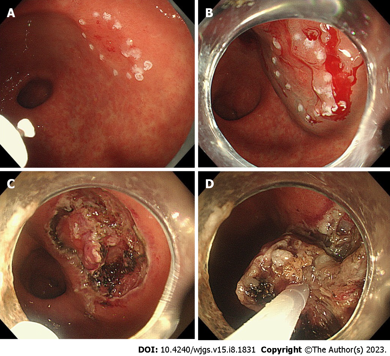
Figure 3 Hybrid endoscopic submucosal dissection process with SOUTEN.
A: A knife attached to the top of the snare is used to mark the lesion; B: A mixture of hyaluronic acid and saline is injected into the submucosa around the lesion; C: A knife attached to the top of the snare is used to dissect the submucosal around the lesion; D: Resection of the lesion with the snare.
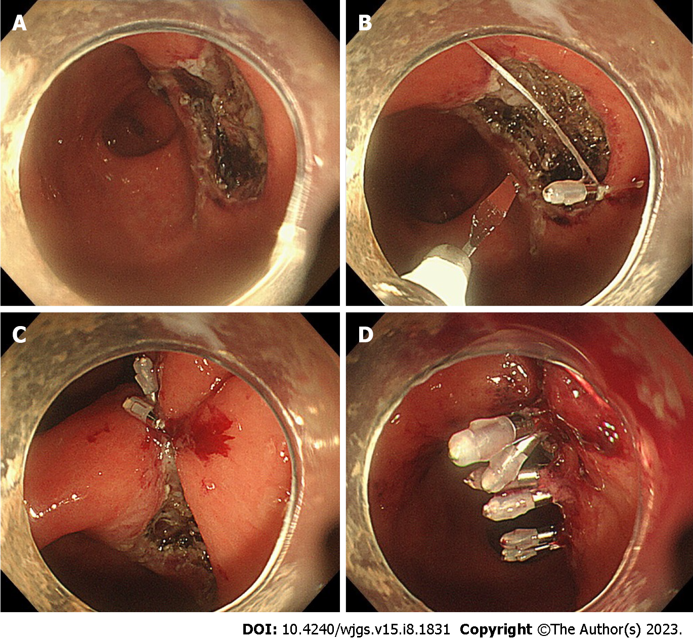
Figure 4 Endoscopic suturing process for a mucosal defect after endoscopic submucosal dissection.
A: Post-endoscopic submucosal dissection mucosal defect in the posterior wall of the gastric antrum; B: A clip with a string attached to the tip is first attached to the anal aspect of the mucosal defect, and an additional clip is attached to the oral aspect of the mucosal defect while sandwiching the string; C: Bring both ends of the mucosa closer by pulling the string out of the mouth; D: Suturing the mucosal defect completely by attaching additional clips to both sides of the mucosal defect and finally burning off the string with the knife of SOUTEN.
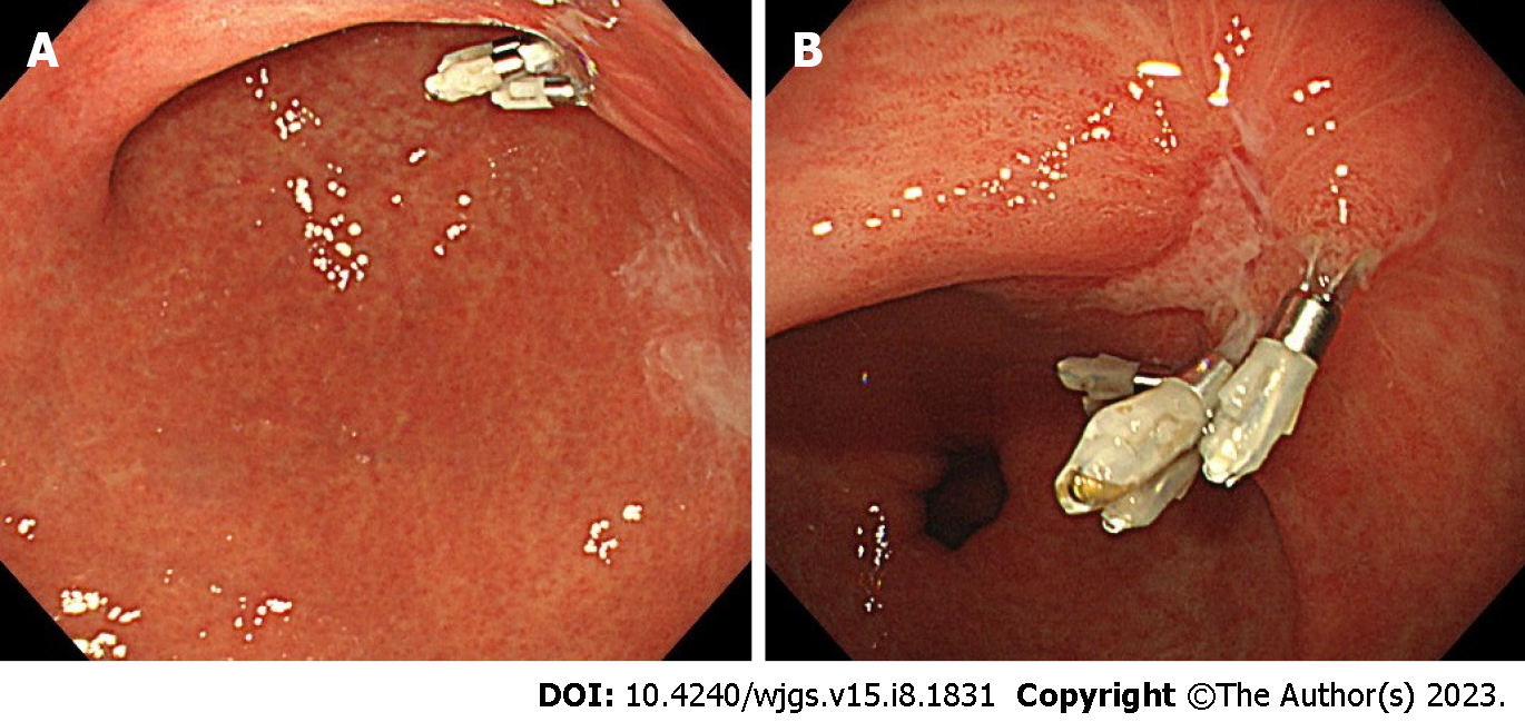
Figure 5 Endoscopic view of the ulcer one month after endoscopic submucosal dissection.
A: Distant view of the ulcer; B: Close-up view of the ulcer.
- Citation: Ito R, Miwa K, Matano Y. Outpatient hybrid endoscopic submucosal dissection with SOUTEN for early gastric cancer, followed by endoscopic suturing of the mucosal defect: A case report. World J Gastrointest Surg 2023; 15(8): 1831-1837
- URL: https://www.wjgnet.com/1948-9366/full/v15/i8/1831.htm
- DOI: https://dx.doi.org/10.4240/wjgs.v15.i8.1831


