Copyright
©The Author(s) 2023.
World J Gastrointest Surg. Aug 27, 2023; 15(8): 1825-1830
Published online Aug 27, 2023. doi: 10.4240/wjgs.v15.i8.1825
Published online Aug 27, 2023. doi: 10.4240/wjgs.v15.i8.1825
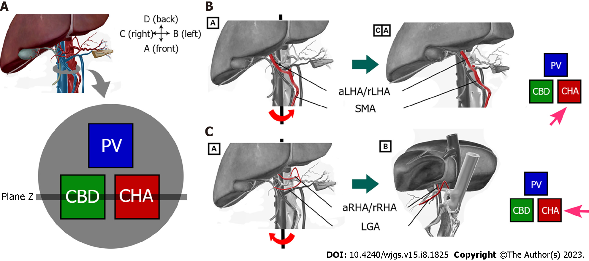
Figure 1 The operating process of the new surgical protocol.
A: Normal hepatic hilar anatomy and cross-sectional diagram. The position relationship between the common bile duct, the portal vein, and the proper hepatic artery is triangular, with the common bile duct located on the right front of the hepatic portal vein and the right side of the proper hepatic artery; B: Dissecting the variant right hepatic artery after rotating the liver counter clockwise; C: Dissecting the variant left hepatic artery after rotating the liver clockwise. aLHA: Accessory left hepatic artery; rLHA: Replaced left hepatic artery; aRHA: Accessory right hepatic artery; rRHA: Replaced right hepatic artery; SMA: Superior mesenteric artery; LGA: Left gastric artery.
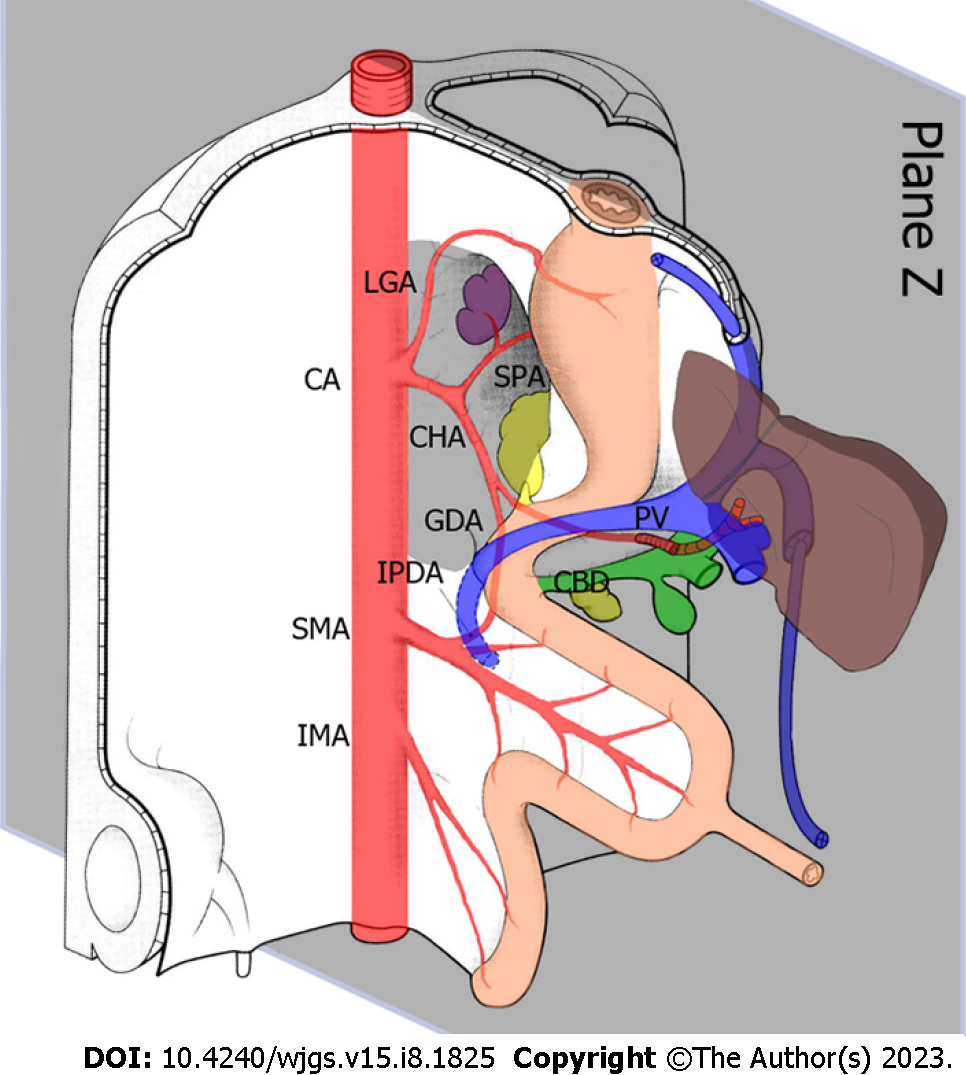
Figure 2 Relationship between the hepatic artery and liver at 5 wk of embryo development.
The grey area represents the position of the ‘Z-plane’, where the variant hepatic artery exists in a two-dimensional plane. CA: Celiac axis; LGA: Left gastric artery; SPA: Splenic artery; CHA: Common hepatic artery; GDA: Gastroduodenal artery; IPDA: Inferior pancreaticoduodenal artery; PV: Portal vein; CBD: Common bile duct; SMA: Superior mesenteric artery; IMA: Inferior mesenteric artery.
- Citation: Zhang HZ, Lu JH, Shi ZY, Guo YR, Shao WH, Meng FX, Zhang R, Zhang AH, Xu J. Donor hepatic artery reconstruction based on human embryology: A case report. World J Gastrointest Surg 2023; 15(8): 1825-1830
- URL: https://www.wjgnet.com/1948-9366/full/v15/i8/1825.htm
- DOI: https://dx.doi.org/10.4240/wjgs.v15.i8.1825


