Copyright
©The Author(s) 2023.
World J Gastrointest Surg. Aug 27, 2023; 15(8): 1819-1824
Published online Aug 27, 2023. doi: 10.4240/wjgs.v15.i8.1819
Published online Aug 27, 2023. doi: 10.4240/wjgs.v15.i8.1819
Figure 1 Abdominal computer tomography showing several heterogenous lesions in the right lobe of the liver.
A: Arterial phase; B: Venous phase; C: Plain scan.
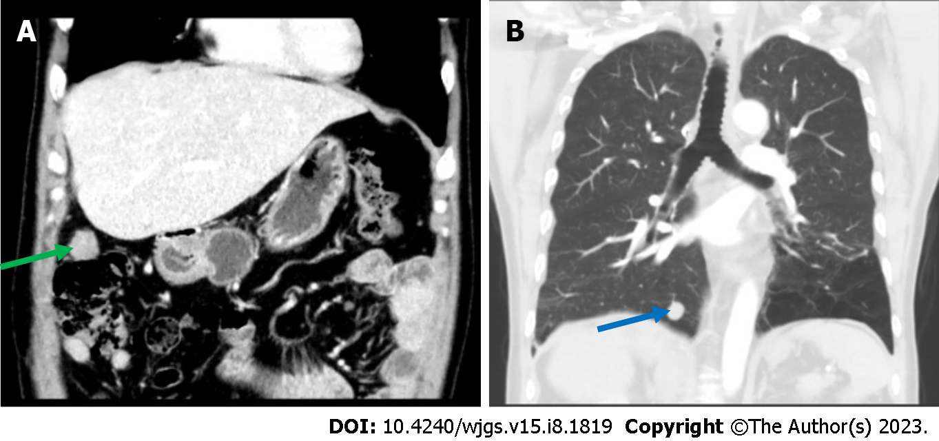
Figure 2 An abdominal computer tomography showed a 2.
0 cm × 1.6 cm nodular soft tissue density lesion in the right upper peritoneum (green arrow), and thoracic computer tomography showed a nodular lesion about 1.6 cm in diameter in the basal segment of the right lower lung (blue arrow). A: An abdominal computer tomography; B: Thoracic computer tomography.
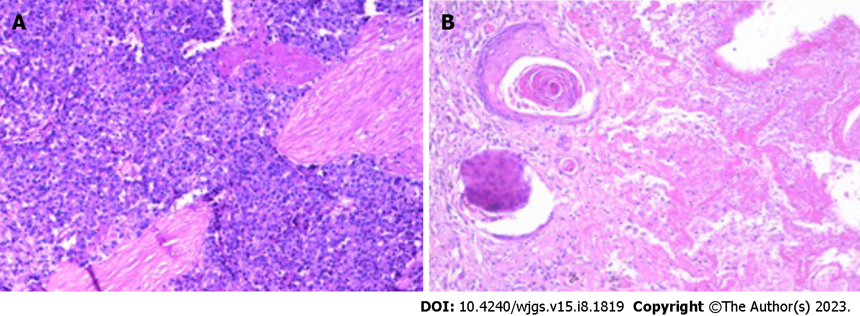
Figure 3 Pathology of the peritoneal mass and pulmonary lesion, which were consistent with metastatic hepatocellular carcinoma.
A: Peritoneal mass; B: Pulmonary lesion.
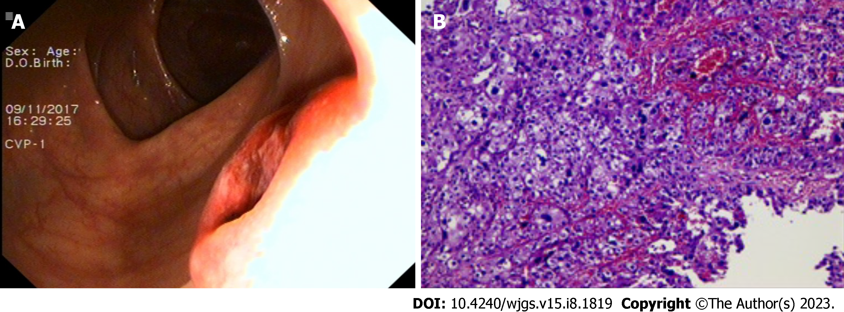
Figure 4 Colonoscopy found a mass in the ascending colon and the biopsy confirmed metastatic hepatocellular carcinoma in the ascending colon.
A: Colonoscopy; B: Biopsy.
- Citation: Gong YQ, Lu TL, Chen CW. Long-term survival of patients with hepatocellular carcinoma with hepatic, pulmonary, peritoneal and rare colon metastasis: A case report. World J Gastrointest Surg 2023; 15(8): 1819-1824
- URL: https://www.wjgnet.com/1948-9366/full/v15/i8/1819.htm
- DOI: https://dx.doi.org/10.4240/wjgs.v15.i8.1819


