Copyright
©The Author(s) 2023.
World J Gastrointest Surg. Jul 27, 2023; 15(7): 1331-1339
Published online Jul 27, 2023. doi: 10.4240/wjgs.v15.i7.1331
Published online Jul 27, 2023. doi: 10.4240/wjgs.v15.i7.1331
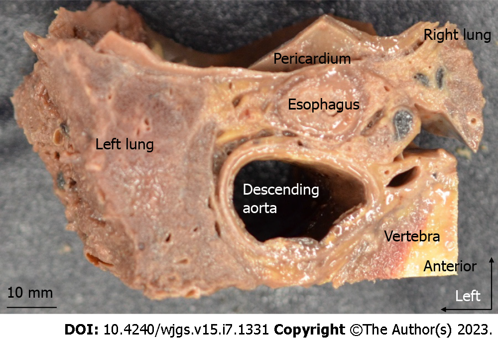
Figure 1 Resected specimen viewed from the cranial side.
The mediastinal organs, vertebrae, and lungs were harvested en-bloc. Organs were dissected at the level of the lower thoracic esophagus. Blocks were then paraffin-embedded and thin sectioned.
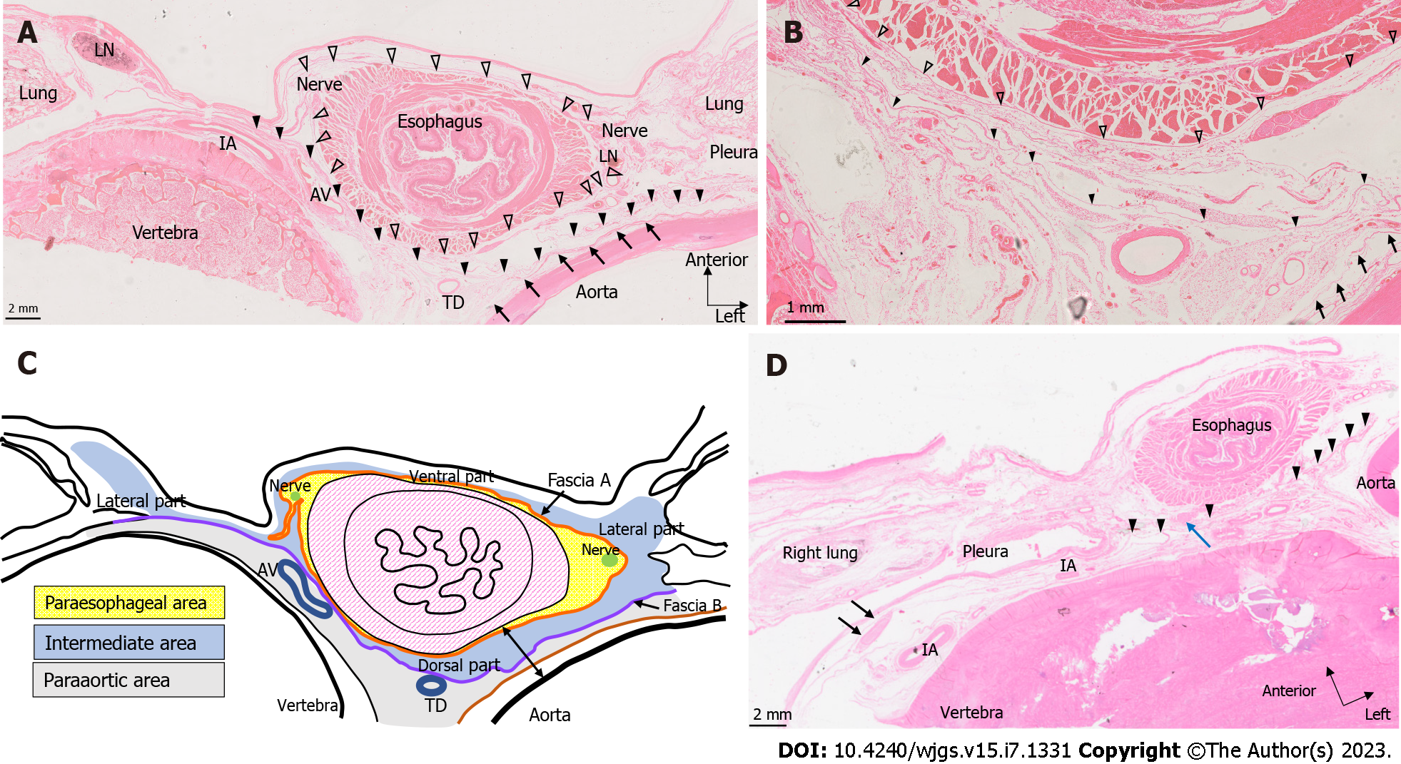
Figure 2 Hematoxylin and eosin-stained tissue sections.
A: Cadaver 3, section 9 mm caudal to the lower pulmonary vein Hematoxylin-Eosin staining (HE-stained). Fascia A closest to the esophageal wall is indicated by white arrowheads. Fascia B on the dorsal side of the esophagus and fascia A is indicated by black arrowheads. The arrows indicate fascia surrounding the descending aorta. In this slide, one small lymph node (LN) is observed within the paraesophageal area on the left side of the esophagus; B: Close-up view of the dorsal side of the esophagus in Figure 2A. Fascia A (white arrowhead) circumferentially surrounds the esophagus and is quite close to the esophageal wall on the dorsal side. Fascia B (black arrowhead) runs dorsal to the esophagus and covers the anterior surface of the thoracic duct and azygos vein. No vessels penetrate these fasciae on the dorsal side; C: Schema created based on cadaver 3 immediately caudal to the inferior pulmonary vein (as in Figure 2B). The yellow area is the paraesophageal area, which contains bilateral vagus nerves and is circumferentially surrounded by fascia A. The. The blue area outside fascia A is the intermediate area, subdivided into left and right lateral, dorsal, and ventral parts. The intermediate area is bounded by fascia B on the dorsal side. The paraaortic area containing peri-thoracic duct LNs extends dorsal to fascia B. The double-ended arrow in the dorsal part demonstrates the distance between the aorta and esophageal wall; D: Cadaver 7, HE-stained. One flat LN is observed on the anterior surface of the vertebral body (black arrow). The arrowheads indicate fascia B. The intercostal artery runs transversely along the curve of the vertebral body. One LN in the dorsal part of the intermediate area is also observed on this slide (blue arrow). AV; Azygos vein; TD; Thoracic duct; LN; Lymph node; IA; Intercostal artery.
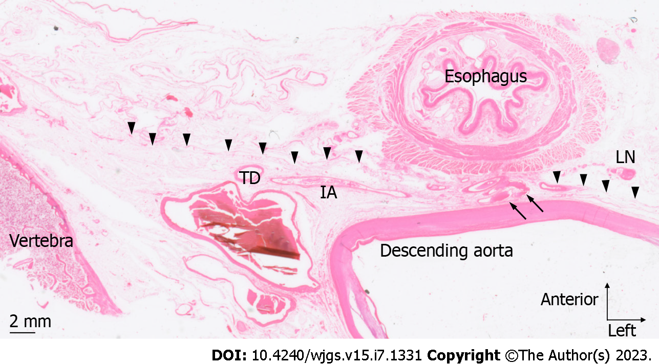
Figure 3 Artery penetrating fascia B.
Cadaver 5, HE-stained section 9 mm cranial to the esophageal hiatus. An artery (arrow) branching from the anterior surface of the descending aorta penetrates fascia B (arrowhead) and runs toward the esophageal wall. No lymph nodes were observed around this artery. TD: Thoracic duct; IA: Intercostal artery; LN: Lymph node.
Figure 4 D2-40-stained tissue sections.
A: D2-40 staining in cadaver 7. The white arrowheads indicate fascia A, and the black arrowheads indicate fascia B. One lymph node (LN) of the lateral part of the intermediate layer is observed in the upper left part of the image. The lymphatic vessels (arrow) near the LNs extend on one side toward the right pleural cavity and lungs, and on the other side toward the ventral side of the esophagus. Furthermore, in this slide, lymphatic vessels in the intermediate area and those in the paraesophageal area were in close proximity; B: Cadaver 3 D2-40-stained section at the lower thoracic esophagus level immediately inferior to the inferior pulmonary vein. The white arrowheads indicate fascia A, and the black arrowheads indicate fascia B. The lateral part of the intermediate area LN is observed in the upper left part of the image, the lymphatic vessels (arrow) near the LNs extend anteriorly into the ventral part of the intermediate area. TD: Thoracic duct; LN: Lymph node; IA: Intercostal artery.
- Citation: Saito T, Muro S, Fujiwara H, Umebayashi Y, Sato Y, Tokunaga M, Akita K, Kinugasa Y. Histological study of the structural layers around the esophagus in the lower mediastinum. World J Gastrointest Surg 2023; 15(7): 1331-1339
- URL: https://www.wjgnet.com/1948-9366/full/v15/i7/1331.htm
- DOI: https://dx.doi.org/10.4240/wjgs.v15.i7.1331


