Copyright
©The Author(s) 2023.
World J Gastrointest Surg. Jun 27, 2023; 15(6): 1247-1255
Published online Jun 27, 2023. doi: 10.4240/wjgs.v15.i6.1247
Published online Jun 27, 2023. doi: 10.4240/wjgs.v15.i6.1247
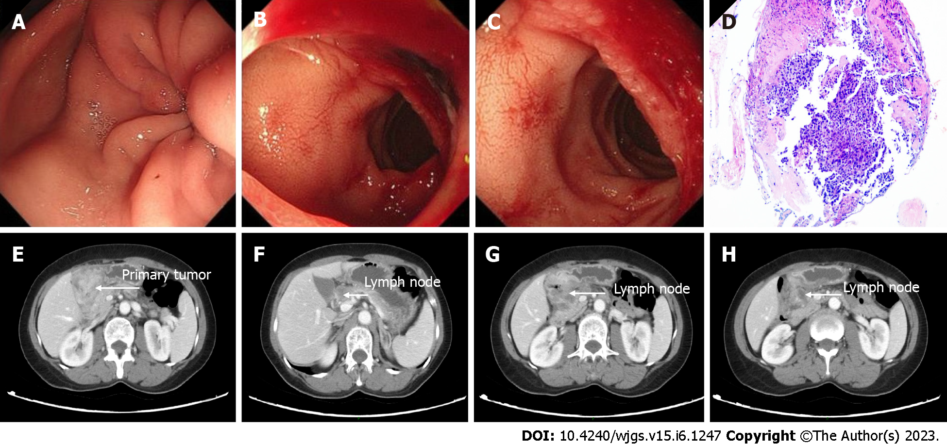
Figure 1 Clinical data of the patient at the initial diagnosis.
A-C: Normal endoscopic showing primary tumor invasion to the antrum (A), pyloric ring (B), and duodenum (C); D: Pathological examination of the endoscopic biopsy revealed a poorly differentiated adenocarcinoma; E-H: Initial computed tomography showing primary tumor invasion to the duodenum (E) and lymph node metastases (F-H).
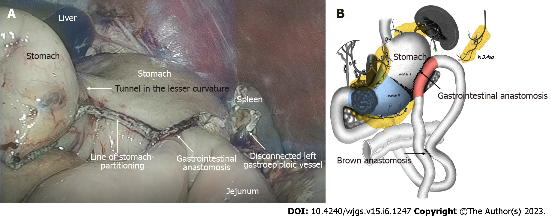
Figure 2 Stomach-partitioning gastrojejunostomy and Braun anastomosis combined with No.
4sb lymph node dissection. A: Intraoperative laparoscopic images; B: Illustration of the modified stomach-partitioning gastrojejunostomy.
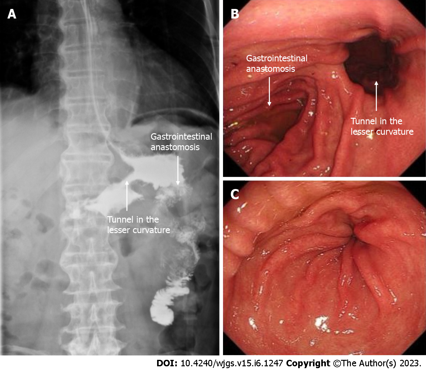
Figure 3 Clinical data of the patient after operation and conversion therapy.
A: Upper digestive tract radiography showed a good passage to the jejunum; B: Endoscopic showing good drainage route in the lesser curvature; C: Endoscopic evidence of good reduction of primary tumor.
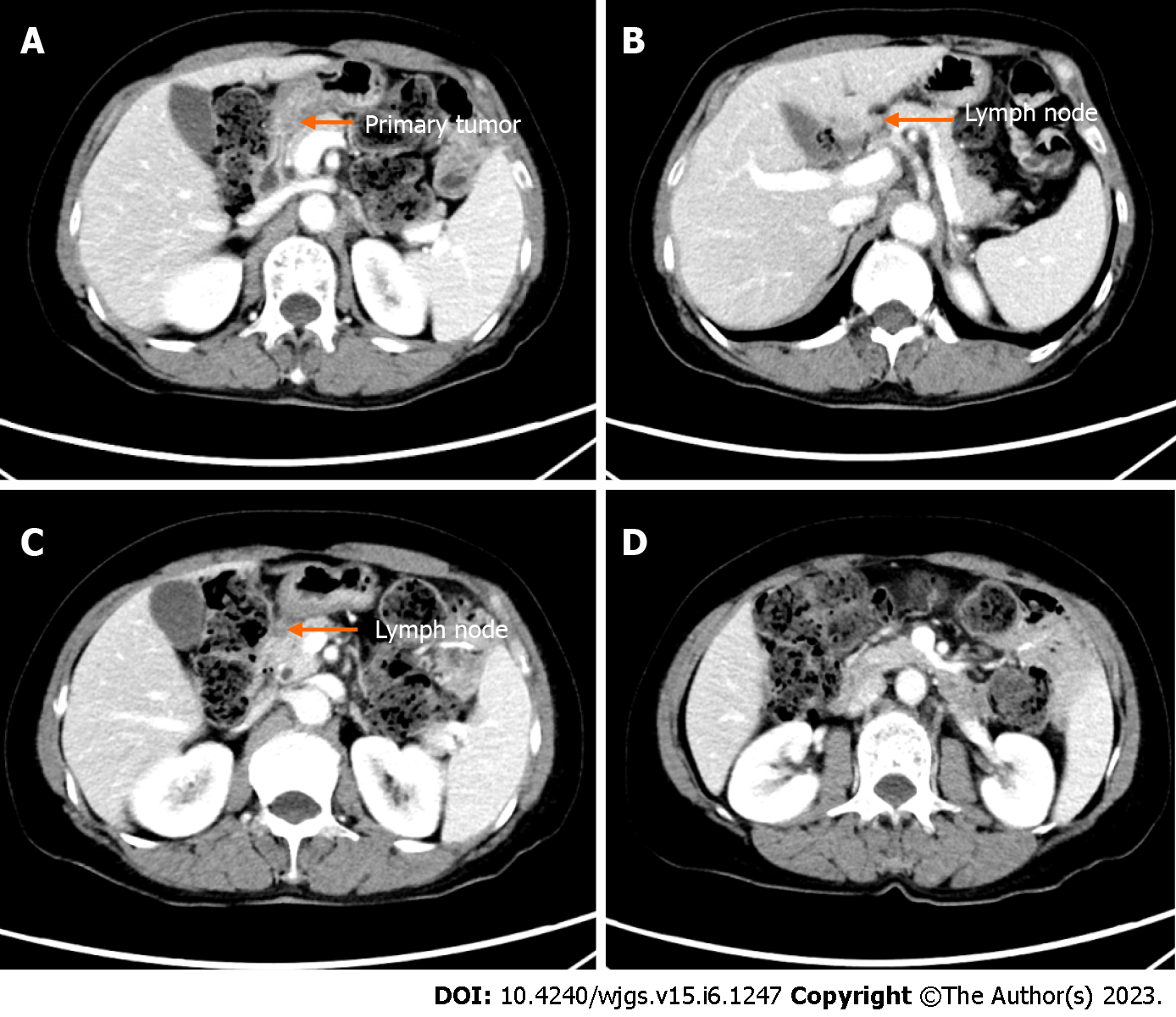
Figure 4 Computed tomography findings after conversion therapy.
A: Primary tumor reduction; B-D: Metastatic lymph node reduction.
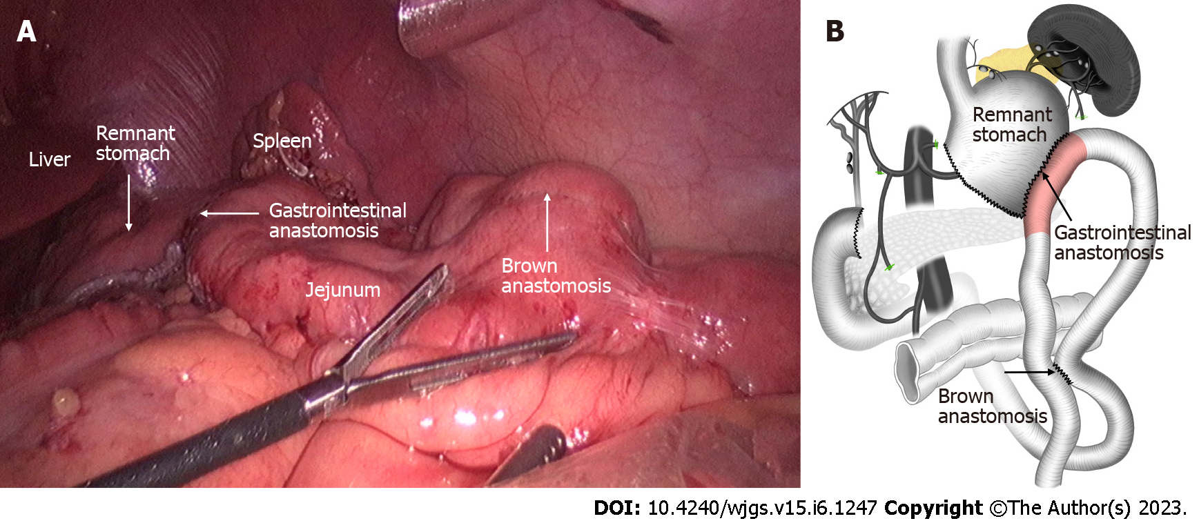
Figure 5 Subtotal gastrectomy with D2 lymph node dissection.
A: Intraoperative laparoscopic images; B: Illustration of the gastrectomy, the anastomoses completed in the previous operation were preserved.
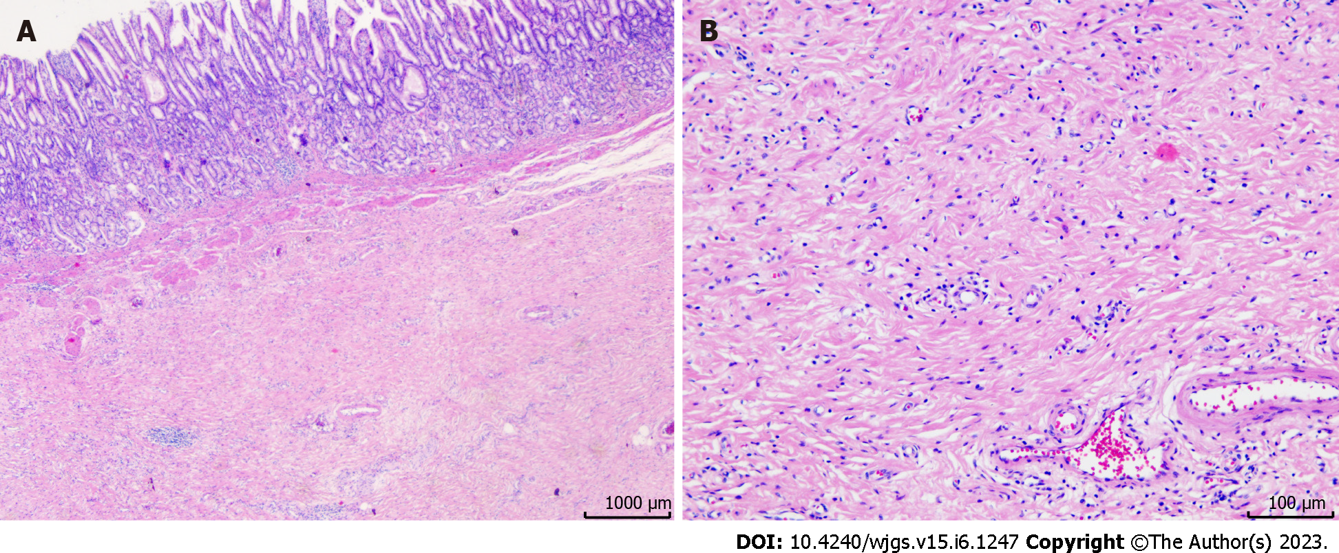
Figure 6 Post-operative pathology showed no residual tumor cells, and a pathological complete response was achieved.
A: Hematoxylin and eosin staining results, × 4; B: Hematoxylin and eosin staining results, × 40.
- Citation: Shao XX, Xu Q, Wang BZ, Tian YT. Modified stomach-partitioning gastrojejunostomy for initially unresectable advanced gastric cancer with outlet obstruction: A case report. World J Gastrointest Surg 2023; 15(6): 1247-1255
- URL: https://www.wjgnet.com/1948-9366/full/v15/i6/1247.htm
- DOI: https://dx.doi.org/10.4240/wjgs.v15.i6.1247


