Copyright
©The Author(s) 2023.
World J Gastrointest Surg. Jun 27, 2023; 15(6): 1224-1231
Published online Jun 27, 2023. doi: 10.4240/wjgs.v15.i6.1224
Published online Jun 27, 2023. doi: 10.4240/wjgs.v15.i6.1224
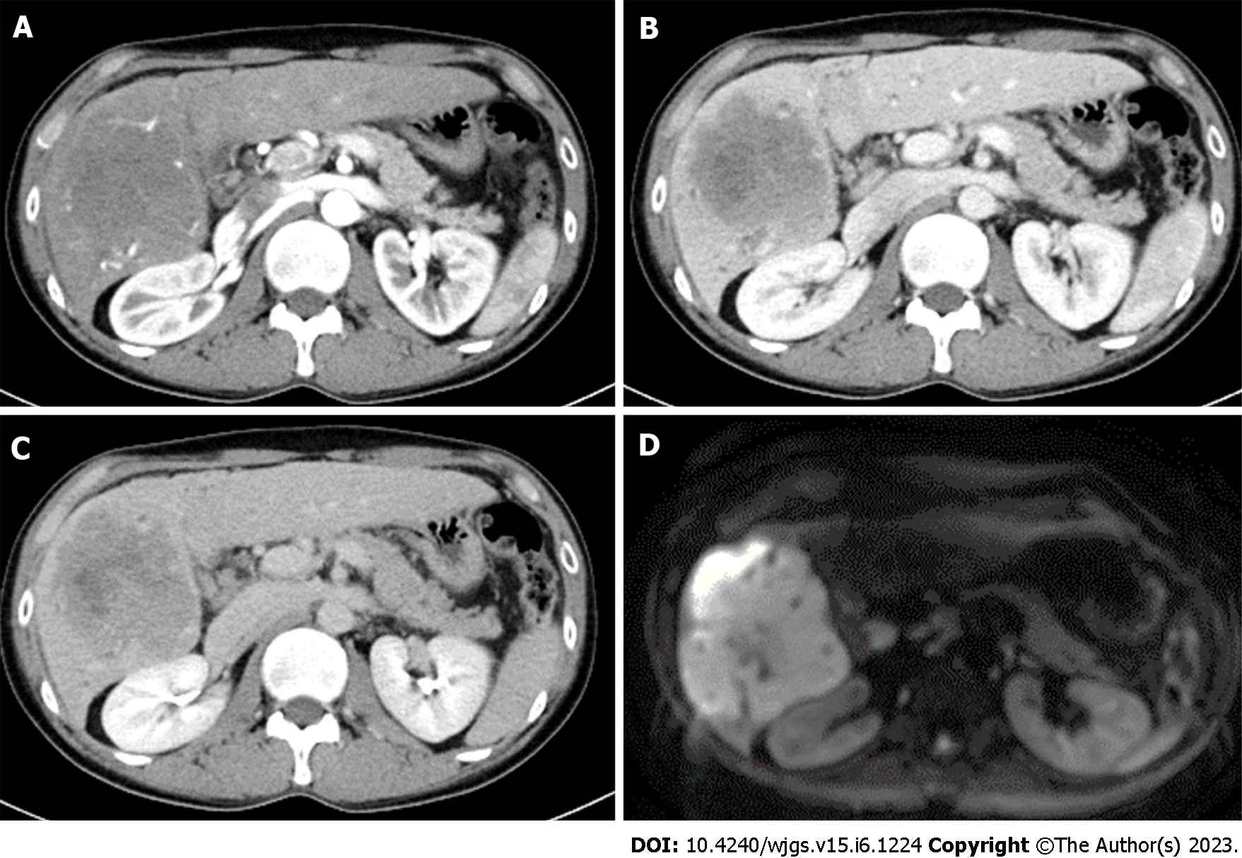
Figure 1 Abdominal contrast-enhanced computed tomography images and magnetic resonance image of case 1.
A: Early arterial phase of computed tomography (CT); B: Portal vein phase of CT; C: Late phase of CT. A massive mass with a major axis of about 10 cm almost occupies the right lobe of the liver S5-6. The mass is gradually stained in a non-uniform ring shape. D: Diffusion weighted image of magnetic resonance image.
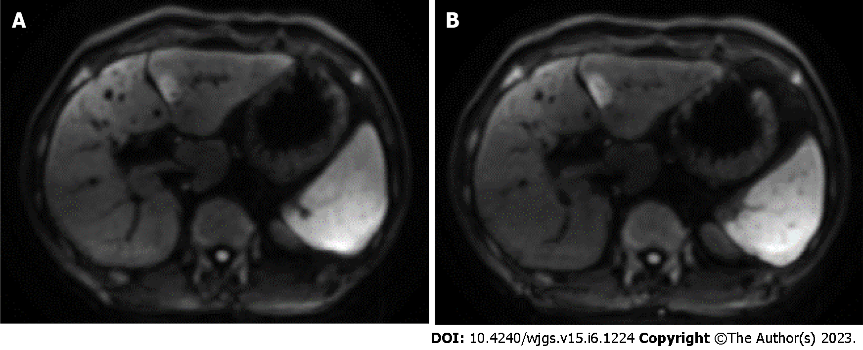
Figure 2 Contrast enhanced magnetic resonance image of case 2.
A: Abdominal contrast-enhanced magnetic resonance image (MRI) diffusion-weighted images show a hyperintensity nodule of about 20 mm in the lateral segment of the left lobe of the liver; B: MRI 4 mo after A. The mass in the lateral section of the left lobe of the liver is 23 mm, which is slightly larger than in the previous image, and the possibility of malignancy could not be ruled out.
Figure 3 Pathological findings of case 1.
A: Macro image shows a large white phyllodes tumor (12.0 cm × 11.8 cm × 10.5 cm); B: Loupe image; C: Micro image shows that the adenocarcinoma is mainly cord-like and has a “partially irregular tubular” to an “obscure tubular” structure. Some areas are accompanied by abundant fibrous stroma.
Figure 4 Pathological findings of case 2.
A: Macro image shows a white to greenish solid mass (17 mm × 16 mm) close to the hepatic sickle mesentery; B: Loupe image; C: Micro image shows arrangement of tubular to papillary, small tubular, and indistinct tubular swelling/infiltration of columnar to rectified atypical cells with mucus. Mucus is found in the glandular cavity with abundant fibrous stroma.
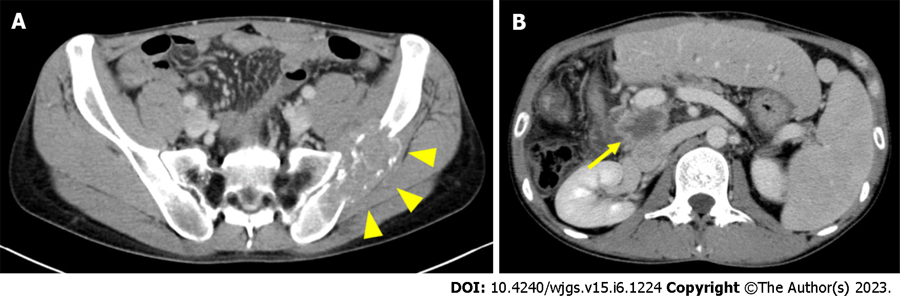
Figure 5 Postoperative contrast enhanced computed tomography of case 1.
A: Left iliac metastasis (arrowhead) is visible 3 mo postoperatively; B: Local recurrence (arrow) is visible on the posterior surface of the portal vein 13 mo postoperatively.
Figure 6 Clinical course of case 1.
RT: Radiation therapy; GEM: Gemcitabine hydrochloride; CDDP: Cisplatin; PTBD: Percutaneous transhepatic biliary drainage.
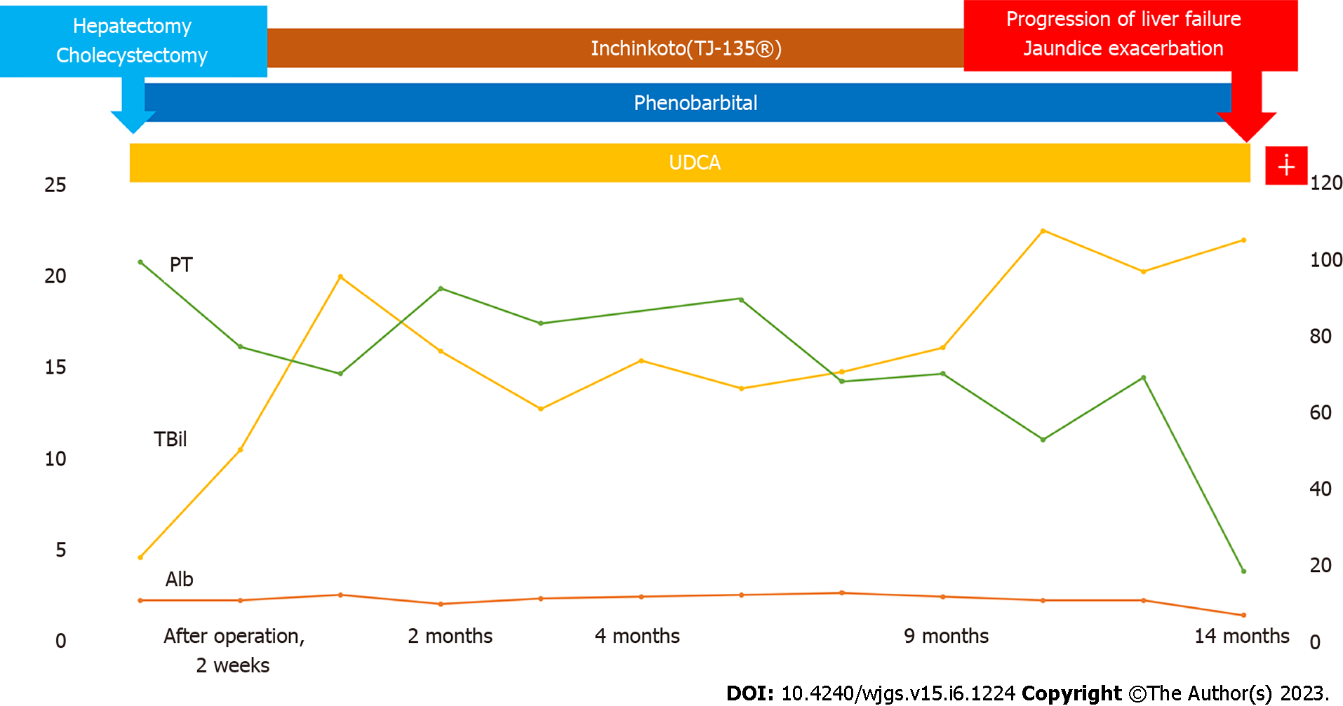
Figure 7 Clinical course of case 2.
UDCA: Ursodeoxycholic acid; PT: Prothrombin time; TBil; Total bilirubin; Alb: Albumin.
- Citation: Miyazu T, Ishida N, Asai Y, Tamura S, Tani S, Yamade M, Iwaizumi M, Hamaya Y, Osawa S, Baba S, Sugimoto K. Intrahepatic cholangiocarcinoma in patients with primary sclerosing cholangitis and ulcerative colitis: Two case reports. World J Gastrointest Surg 2023; 15(6): 1224-1231
- URL: https://www.wjgnet.com/1948-9366/full/v15/i6/1224.htm
- DOI: https://dx.doi.org/10.4240/wjgs.v15.i6.1224


