Copyright
©The Author(s) 2023.
World J Gastrointest Surg. Jun 27, 2023; 15(6): 1211-1215
Published online Jun 27, 2023. doi: 10.4240/wjgs.v15.i6.1211
Published online Jun 27, 2023. doi: 10.4240/wjgs.v15.i6.1211
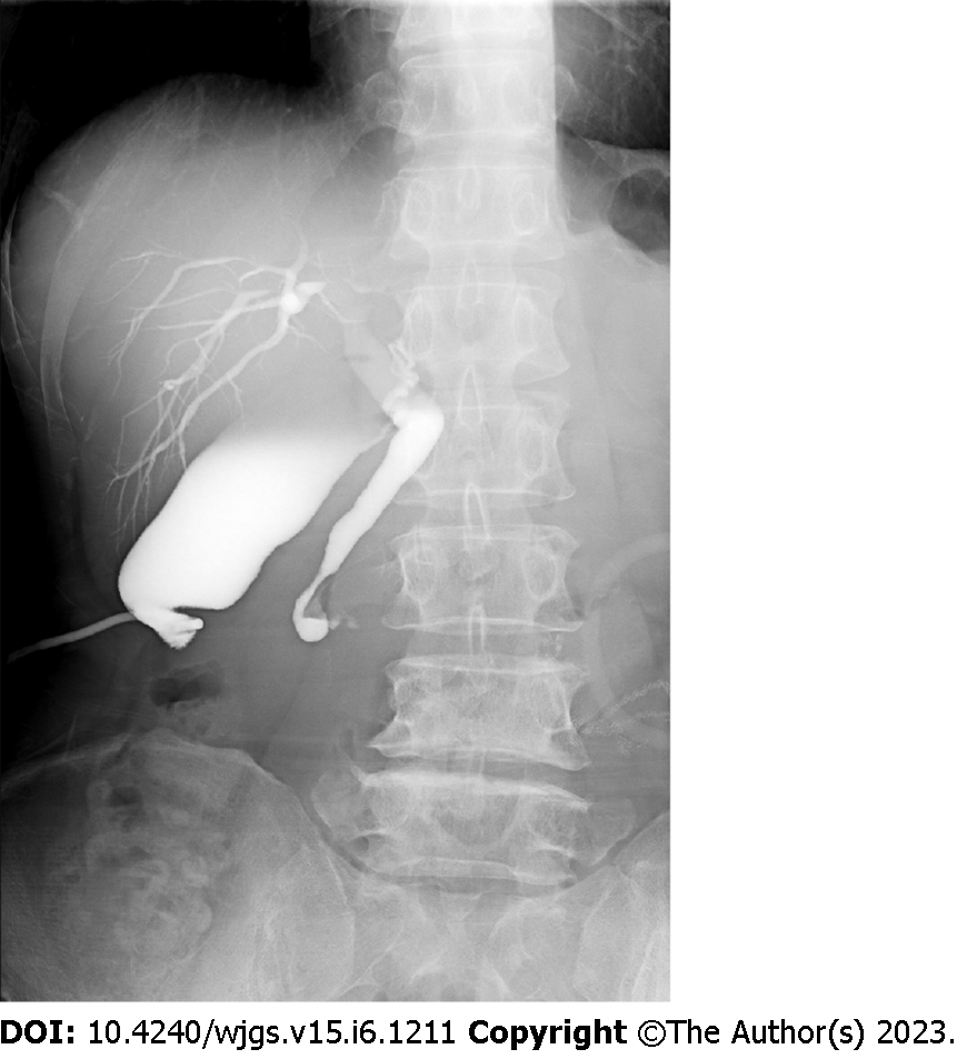
Figure 1 Transcholecystostomy imaging showing slight common bile duct dilation.
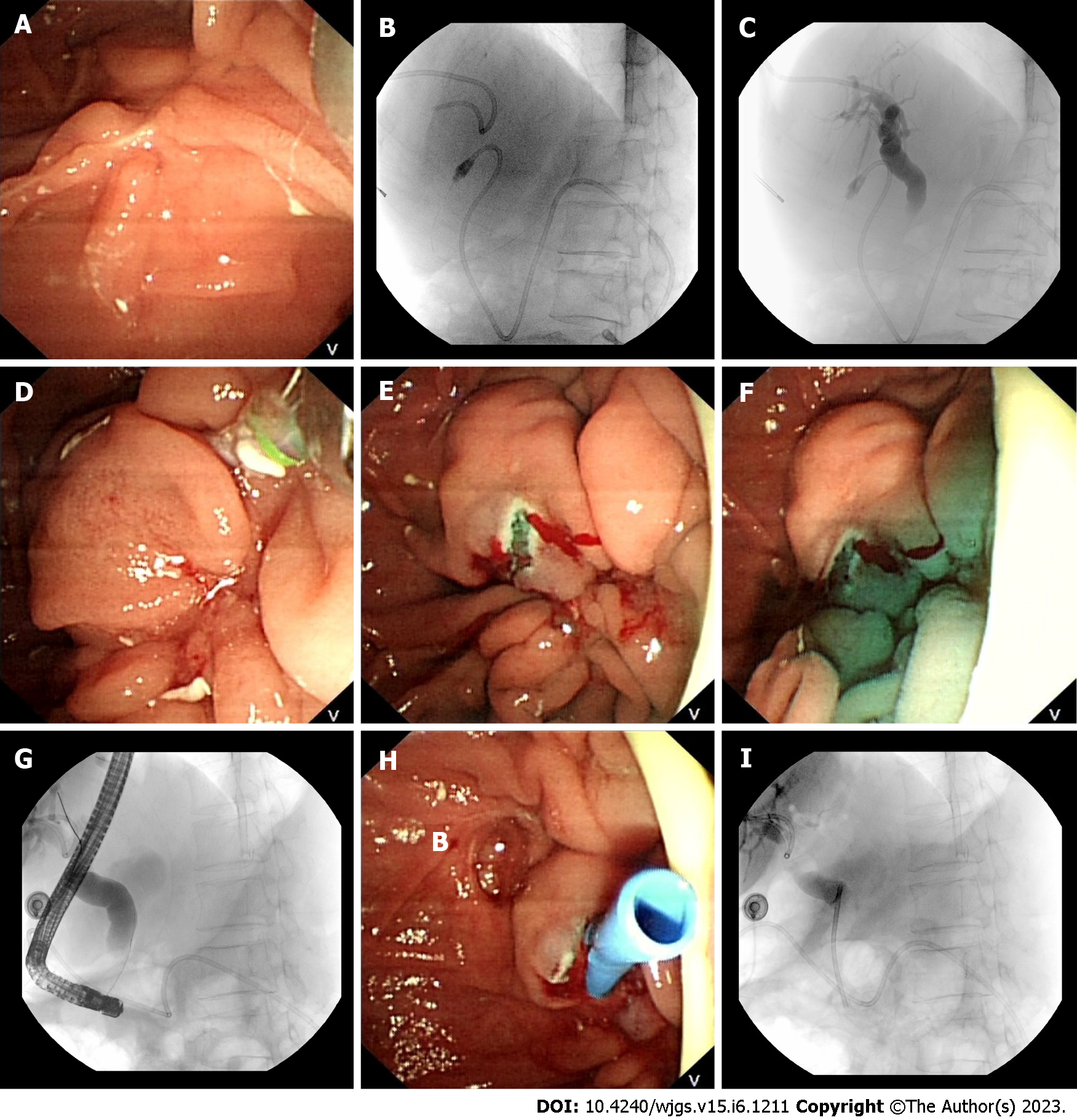
Figure 2 Treatment.
A: Repeated failed attempts to identify the duodenal papilla, a small amount of contrast agent entered the duodenum; B and C: Repeated attempts at guidewire insertion through the percutaneous transhepatic cholangial drainage (PTCD) tube failed; D: mixture of ioversol and methylene blue was injected via the PTCD tube; E: Dual-knife was used for layer-by-layer resection. Pale blue-colored protrusions, which were considered to be the intramural common bile duct, can be seen at the duodenal scar; F: A large amount of methylene blue flowed out after dual-knife resection; G: Common bile duct dilation was observed on endoscopic retrograde cholangiopancreatography imaging; H: Insertion of an 8.5 Fr × 5.0 cm plastic stent; I: A large amount of ioversol and methylene can be seen flowing out.
- Citation: Tang BX, Li XL, Wei N, Tao T. Percutaneous transhepatic cholangial drainage-guided methylene blue for fistulotomy using dual-knife for bile duct intubation: A case report. World J Gastrointest Surg 2023; 15(6): 1211-1215
- URL: https://www.wjgnet.com/1948-9366/full/v15/i6/1211.htm
- DOI: https://dx.doi.org/10.4240/wjgs.v15.i6.1211


