Copyright
©The Author(s) 2023.
World J Gastrointest Surg. Jun 27, 2023; 15(6): 1056-1067
Published online Jun 27, 2023. doi: 10.4240/wjgs.v15.i6.1056
Published online Jun 27, 2023. doi: 10.4240/wjgs.v15.i6.1056
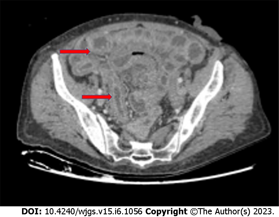
Figure 1 Chronic radiation enteritis in a 62-year-old woman with anal cancer.
Red arrows indicate regions where radiation enteritis is most evident (personal observation).
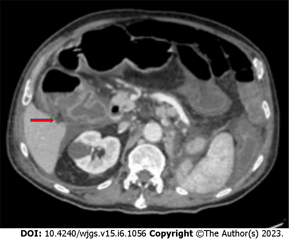
Figure 2 Clostridium difficile colitis (red arrow) in a 78-year-old man with a malignant tumor of the lung treated with cyclophosphamide.
(personal observation).
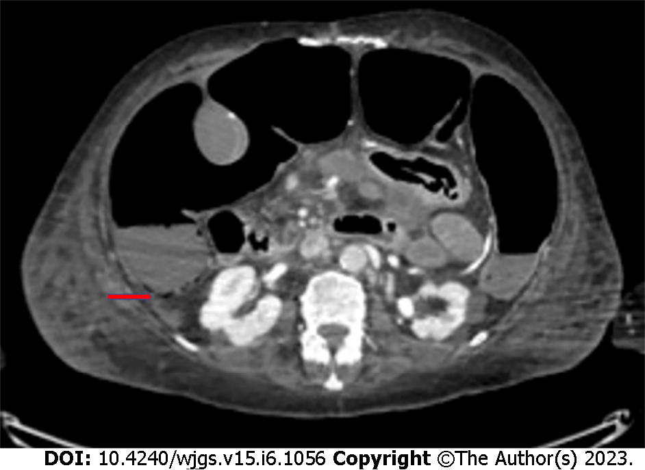
Figure 3 Bevacizumab-related intestinal pneumatosis with right colon ischemia in a 69-year-old woman being treated for breast cancer.
Red arrows indicate regions where pneumatosis is most evident (personal observation).
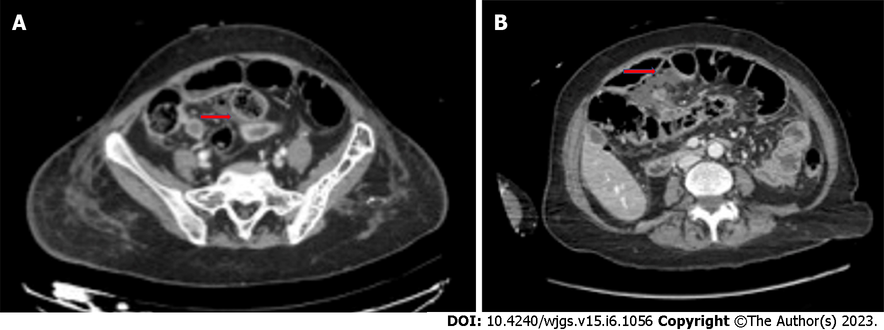
Figure 4 Computed tomography images.
A: Bevacizumab-related small bowel perforation in a 49-year-old female patient with breast cancer (red arrow); B: Bevacizumab-related late anastomotic leakage (red arrow) in a 72-year-old female colon cancer patient (personal observation).
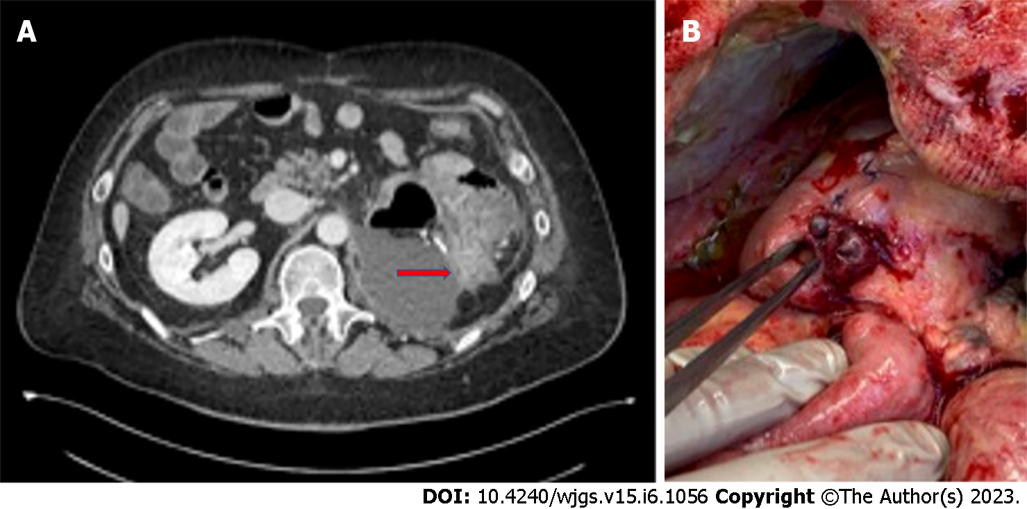
Figure 5 Computed tomography and intraoperative findings.
A: Computed tomography scan of bowel perforation (red arrow) in a 56-year-old male patient undergoing molecular targeted therapy with capozatinib for metastatic renal cell carcinoma; B: Intraoperative findings in the same clinical case (personal observation).
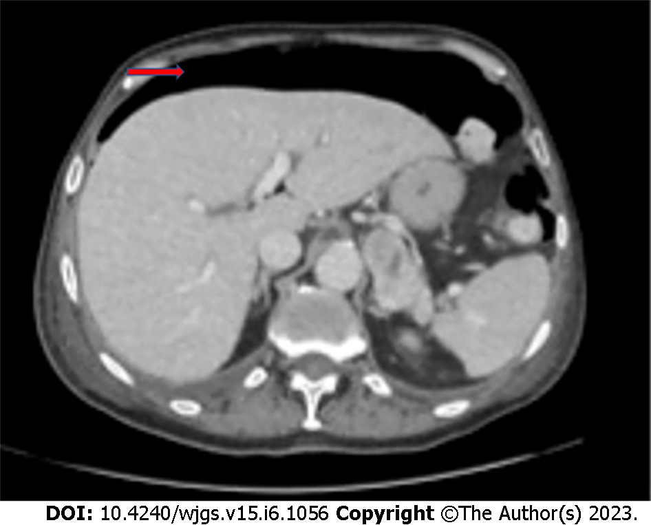
Figure 6 Bowel perforation in a 73-year-old male lung cancer patient undergoing immunotherapy with atezolizumab.
The red arrow indicates subdiaphragmatic free air (personal observation).
- Citation: Fico V, Altieri G, Di Grezia M, Bianchi V, Chiarello MM, Pepe G, Tropeano G, Brisinda G. Surgical complications of oncological treatments: A narrative review. World J Gastrointest Surg 2023; 15(6): 1056-1067
- URL: https://www.wjgnet.com/1948-9366/full/v15/i6/1056.htm
- DOI: https://dx.doi.org/10.4240/wjgs.v15.i6.1056


