Copyright
©The Author(s) 2023.
World J Gastrointest Surg. May 27, 2023; 15(5): 825-833
Published online May 27, 2023. doi: 10.4240/wjgs.v15.i5.825
Published online May 27, 2023. doi: 10.4240/wjgs.v15.i5.825
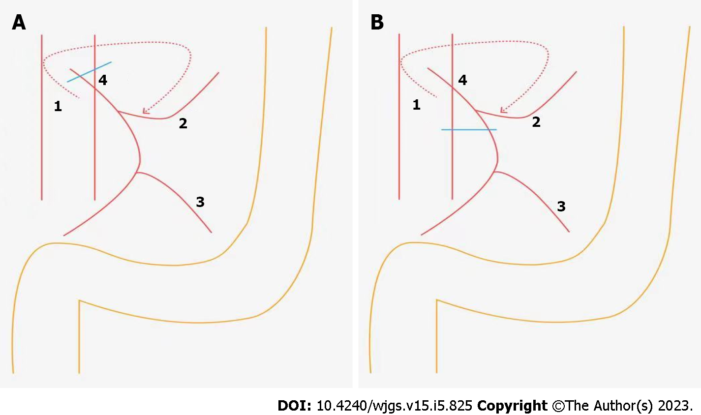
Figure 1 The diagram of ligation site of the inferior mesenteric artery in colorectal cancer surgery.
A: High tie; B: Low tie. 1: Inferior mesenteric artery; 2: Left colic artery; 3: The first branch of the sigmoid artery; 4: Lymph node around the origin of the inferior mesenteric artery.
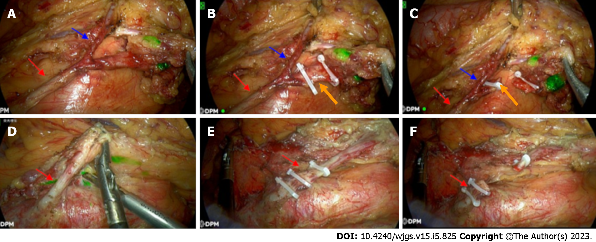
Figure 2 The intraoperative photographs of the ligation site of the inferior mesenteric artery in colorectal cancer surgery.
A-C: Low ligation combined with lymph node dissection around the origin of the inferior mesenteric artery (low tie with lymph node); D-F: High ligation combined with lymph node dissection around the origin of the inferior mesenteric artery (high tie with LND). Red arrows: The route of the inferior mesenteric artery; Blue arrows: Left colic artery; Orange arrows: Superior rectal artery.
- Citation: Liu FC, Song JN, Yang YC, Zhang ZT. Preservation of left colic artery in laparoscopic colorectal operation: The benefit challenge. World J Gastrointest Surg 2023; 15(5): 825-833
- URL: https://www.wjgnet.com/1948-9366/full/v15/i5/825.htm
- DOI: https://dx.doi.org/10.4240/wjgs.v15.i5.825


