Copyright
©The Author(s) 2023.
World J Gastrointest Surg. May 27, 2023; 15(5): 1000-1006
Published online May 27, 2023. doi: 10.4240/wjgs.v15.i5.1000
Published online May 27, 2023. doi: 10.4240/wjgs.v15.i5.1000
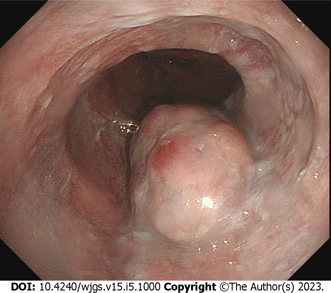
Figure 1 Endoscopic findings.
One 35-mm submucosal tumor was located in the lower thoracic esophagus, with a central depression with a reddish appearance.
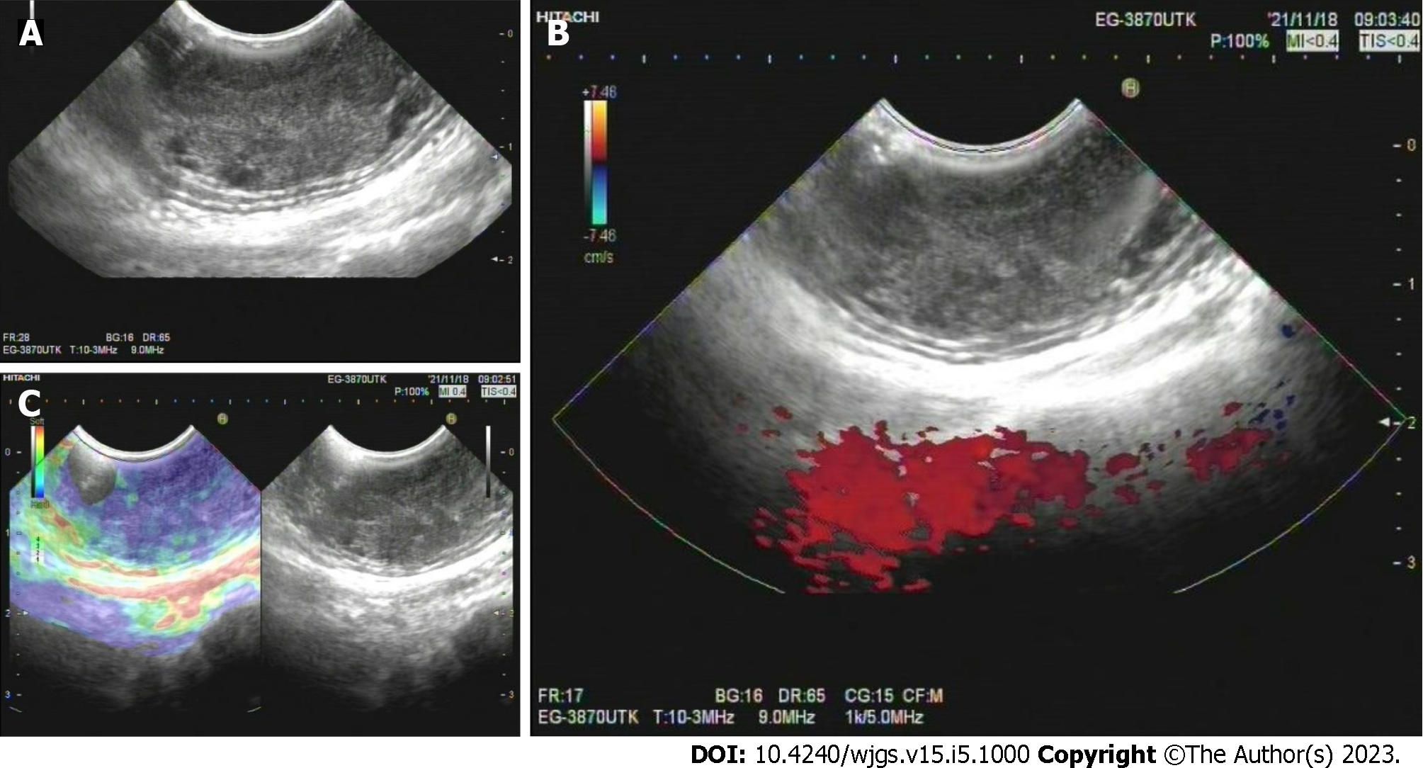
Figure 2 Endoscopic ultrasound findings.
A: A well-defined heterogeneous, hypoechoic lesion with scattered small anechoic areas in the third layer; B: No vascular flow within the lesion; C: A hard mass revealed by elastography.
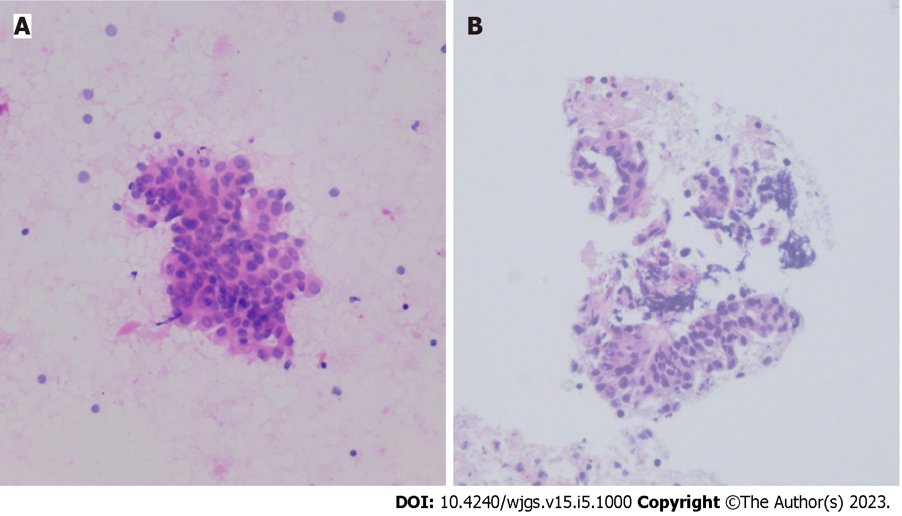
Figure 3 Endoscopic ultrasound - fine-needle aspiration findings.
A: Cytological analysis; B: Histological analysis.
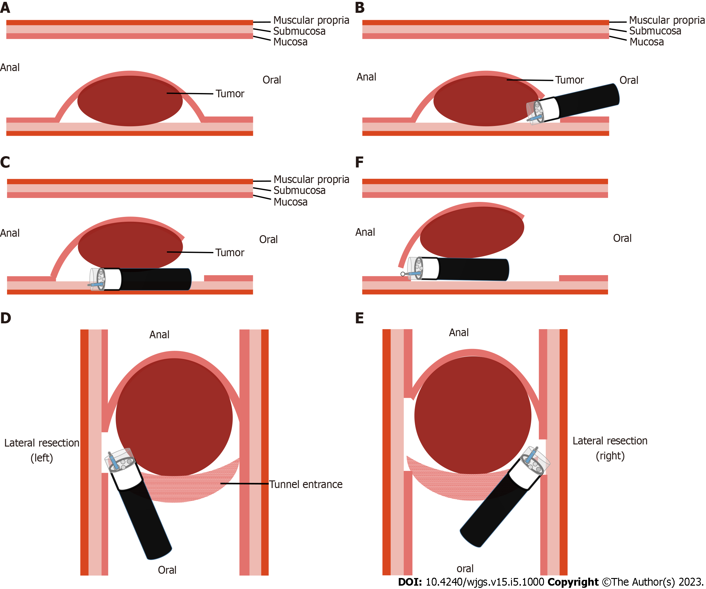
Figure 4 Schema of modified endoscopic submucosal tunnel dissection.
A: Endoscopic submucosal tunnel dissection located in the submucosal layer; B: Oral end of the involved mucosa cut transversely; C: Submucosal tunnel created from the proximal end to the distal end; D: One lateral mucosa resected; E: The other lateral mucosa resected; F: Anal end of the involved mucosa resected.
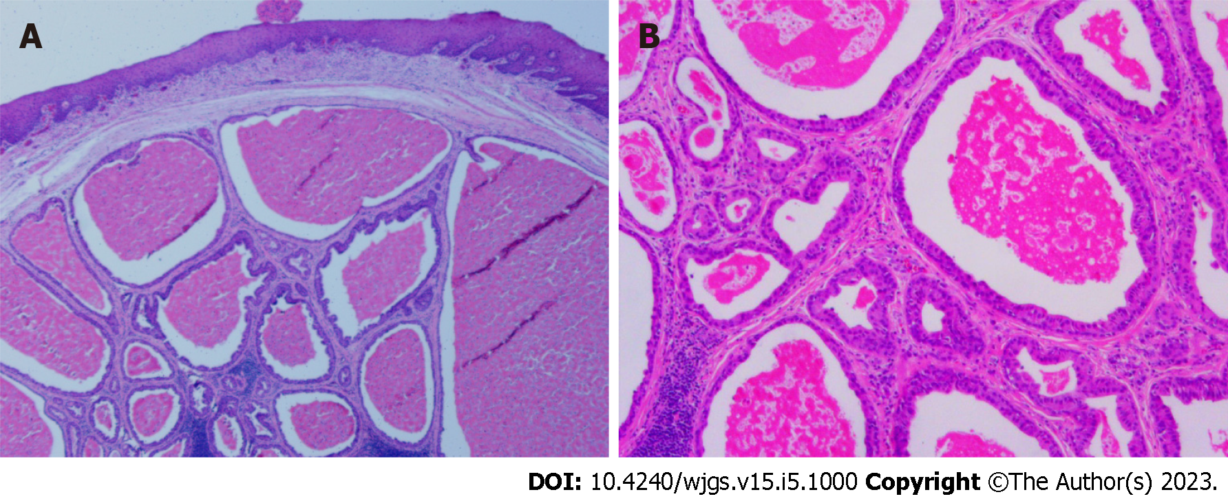
Figure 5 Post-Endoscopic submucosal tunnel dissection pathology.
A: A cystic pattern with distinct 2-cell layers; B: Eosinophilic inner luminal cells.
- Citation: Chen SY, Xie ZF, Jiang Y, Lin J, Shi H. Modified endoscopic submucosal tunnel dissection for large esophageal submucosal gland duct adenoma: A case report. World J Gastrointest Surg 2023; 15(5): 1000-1006
- URL: https://www.wjgnet.com/1948-9366/full/v15/i5/1000.htm
- DOI: https://dx.doi.org/10.4240/wjgs.v15.i5.1000


