Copyright
©The Author(s) 2023.
World J Gastrointest Surg. Apr 27, 2023; 15(4): 674-686
Published online Apr 27, 2023. doi: 10.4240/wjgs.v15.i4.674
Published online Apr 27, 2023. doi: 10.4240/wjgs.v15.i4.674
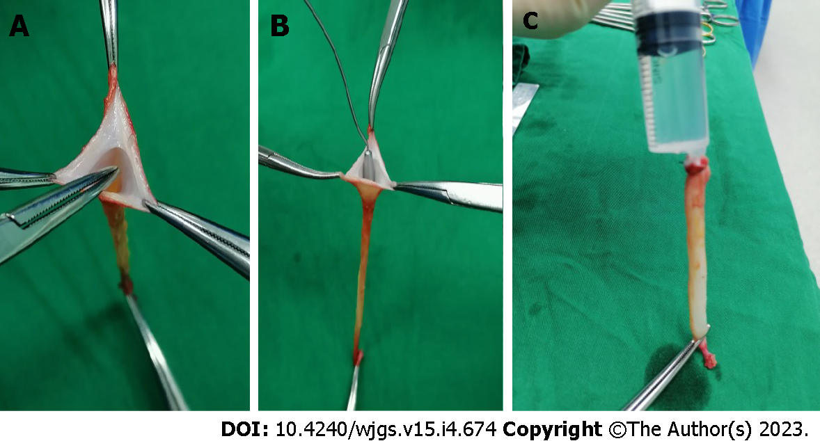
Figure 1 Ligamentum teres hepatis recanalization.
A and B: The remnant lumen of the ligamentum teres hepatis was identified and recanalized using a mosquito clamp (A) and a 3 mm probe (B); C: A bolus of normal saline was injected into the lumen from the proximal end to further enlarge the lumen.
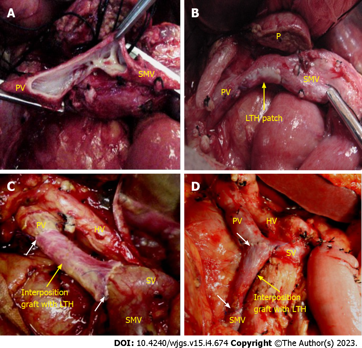
Figure 2 Vascular reconstruction.
A: Tangential resection of the involved vein with the preservation of portal vein (PV)-splenic vein (SV)-superior mesenteric vein (SMV) confluence; B: Vein reconstruction with ligamentum teres hepatis (LTH) patch; C: PV reconstruction with an interposition LTH graft with the preservation of SV-SMV confluence; D: Repair of the SMV with an interposition LTH graft with the preservation of PV-SV confluence. The white arrows indicate the anastomosis line. LTH: Ligamentum teres hepatis; PV: Portal vein; SMV: Superior mesenteric vein; SV: Splenic vein; P: Pancreas; HA: Hepatic artery.
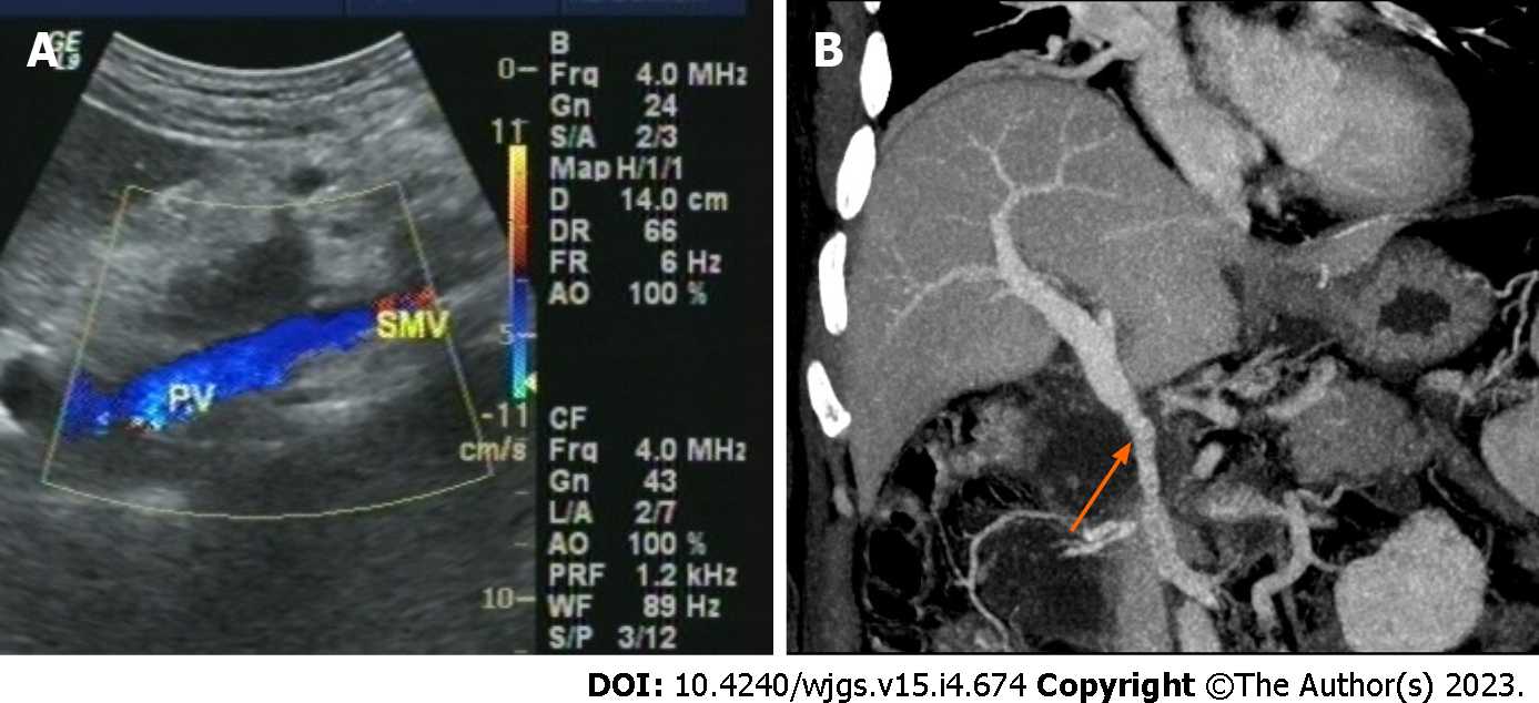
Figure 3 Images of the reconstructed portal vein and/or superior mesenteric vein.
A: Doppler ultrasound showed a patent vascular lumen; B: Enhanced computed tomography scan revealed a patent vascular lumen (orange arrow). PV: Portal vein; SMV: Superior mesenteric vein.
Figure 4 Distribution and function of endothelial cells and relative content of collagen fibers, elastic fibers and smooth muscle in ligamentum teres hepatis.
A: The ligamentum teres hepatis (LTH) lumen (× 100 magnification); B: Endothelial cells (black arrow, × 400 magnification); C: LTH after Verhoeff-Van Gieson (VVG) staining (× 400 magnification); D: The portal vein (PV) after VVG staining. Elastic fibers (red), collagen fibers (black) and smooth muscle (yellow) (× 400 magnification); E: CD34 expression in LTH endothelial cells (× 400 magnification); F: CD34 expression in PV endothelial cells (× 400 magnification); G: Factor VIII-related antigen (FVIIIAg) expression in LTH endothelial cells (× 400 magnification); H: FVIIIAg expression in PV endothelial cells (× 400 magnification); I: Endothelial nitric oxide synthase (eNOS) expression in LTH endothelial cells (× 400 magnification); J: eNOS expression in PV endothelial cells (× 400 magnification); K: Tissue type plasminogen activator (t-PA) expression in LTH endothelial cells (× 400 magnification); L: Tissue type plasminogen activator expression in PV endothelial cells (× 400 magnification).
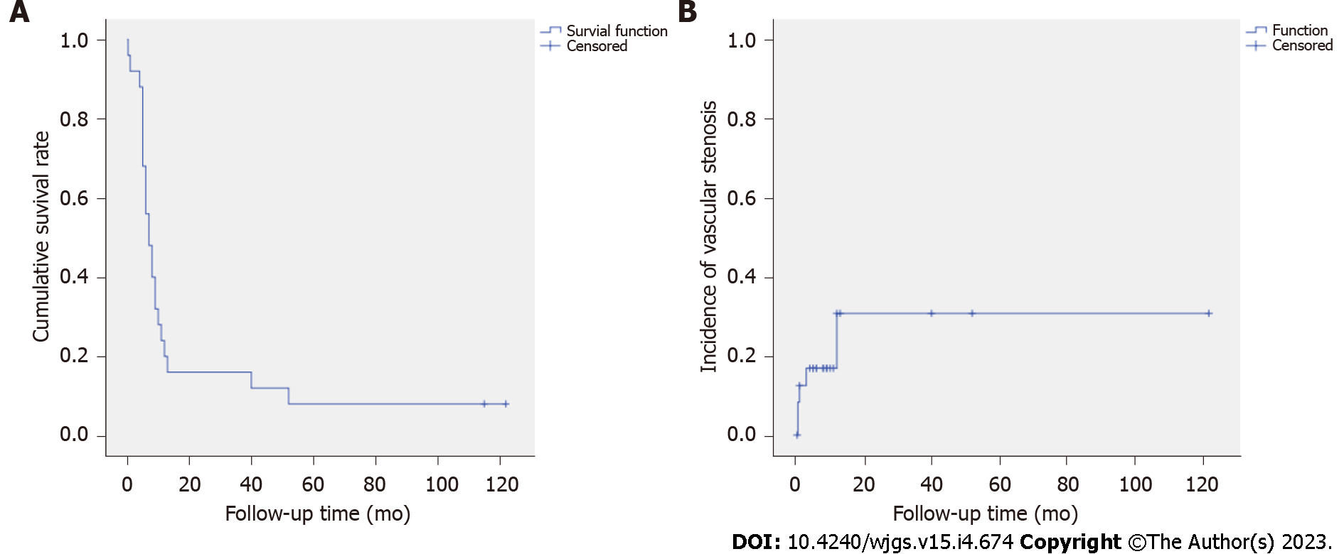
Figure 5 Postoperative cumulative survival and vascular stenosis rate curve.
A: The cumulative survival curve of the 26 patients undergoing pancreatoduodenectomy with venous resection and reconstruction; B: The vascular stenosis rate curve of the 26 patients undergoing portal vein and/or superior mesenteric vein reconstruction with ligamentum teres hepatis.
- Citation: Zhu WT, Wang HT, Guan QH, Zhang F, Zhang CX, Hu FA, Zhao BL, Zhou L, Wei Q, Ji HB, Fu TL, Zhang XY, Wang RT, Chen QP. Ligamentum teres hepatis as a graft for portal and/or superior mesenteric vein reconstruction: From bench to bedside. World J Gastrointest Surg 2023; 15(4): 674-686
- URL: https://www.wjgnet.com/1948-9366/full/v15/i4/674.htm
- DOI: https://dx.doi.org/10.4240/wjgs.v15.i4.674


