Copyright
©The Author(s) 2023.
World J Gastrointest Surg. Apr 27, 2023; 15(4): 534-543
Published online Apr 27, 2023. doi: 10.4240/wjgs.v15.i4.534
Published online Apr 27, 2023. doi: 10.4240/wjgs.v15.i4.534
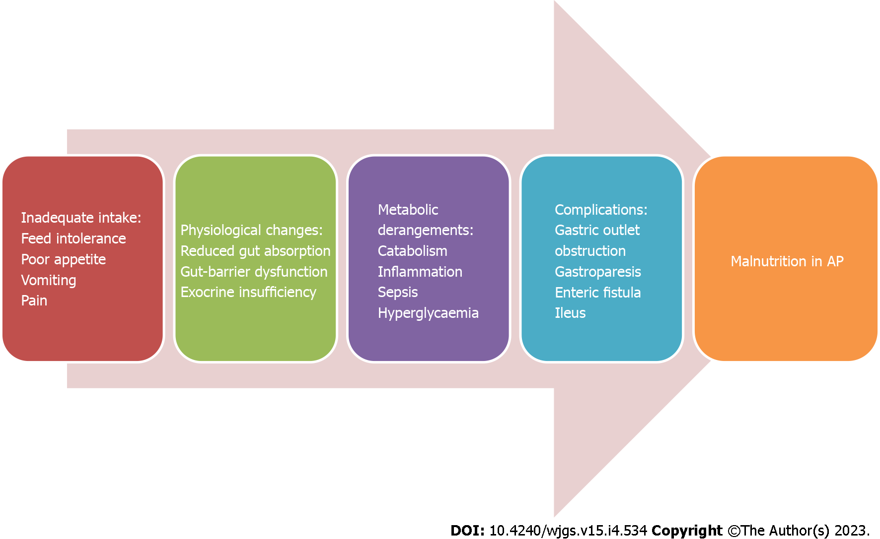
Figure 1 Various probable causes of malnutrition during the course of acute pancreatitis.
AP: Acute pancreatitis.
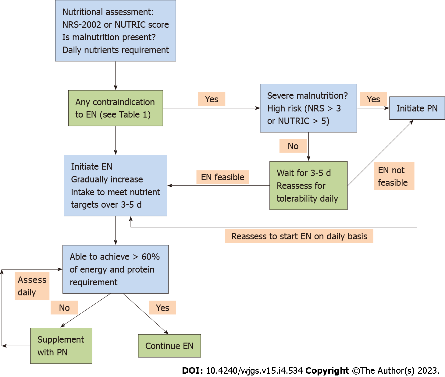
Figure 2 Flowchart of the initiation of nutrition in patients with acute pancreatitis.
EN: Enteral nutrition; NRS: Nutritional risk screening; NUTRIC: Nutrition risk in the critically ill; PN: Parenteral nutrition.
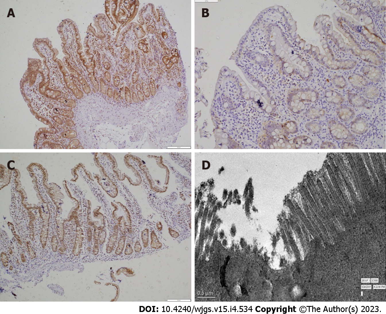
Figure 3 Gut barrier dysfunction and its restoration after enteral nutrition.
A: Duodenal biopsy from control shows intact claudin-3 positivity on immunohistochemistry in both villi and crypts throughout the mucosa (× 200); B: Biopsy taken from acute pancreatitis (AP) shows loss of claudin-3 positivity in the duodenal villi and crypts (× 200); C: Biopsy taken from AP post-enteral nutrition shows positivity (significant improvement) in the duodenal villi and crypts (× 200). Ultrastructural changes in duodenal epithelia of patients with AP on electron microscopy show disordered microvilli.
- Citation: Gopi S, Saraya A, Gunjan D. Nutrition in acute pancreatitis. World J Gastrointest Surg 2023; 15(4): 534-543
- URL: https://www.wjgnet.com/1948-9366/full/v15/i4/534.htm
- DOI: https://dx.doi.org/10.4240/wjgs.v15.i4.534


