Copyright
©The Author(s) 2023.
World J Gastrointest Surg. Dec 27, 2023; 15(12): 2919-2925
Published online Dec 27, 2023. doi: 10.4240/wjgs.v15.i12.2919
Published online Dec 27, 2023. doi: 10.4240/wjgs.v15.i12.2919
Figure 1
Preoperative X-ray film using iohexol as the contrast agent.
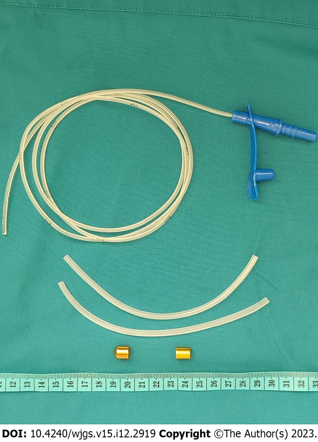
Figure 2
Magnetic compression system.
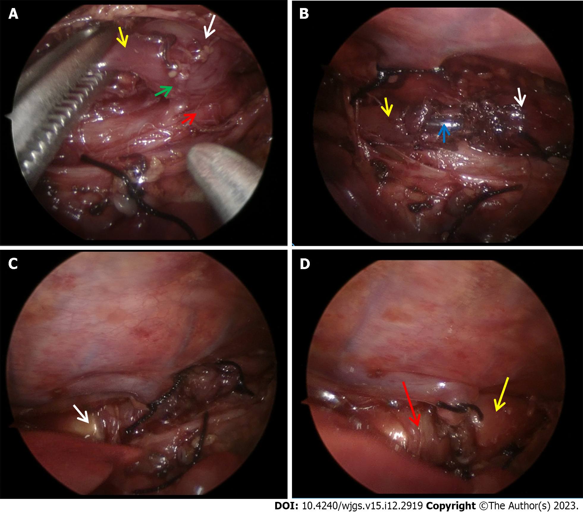
Figure 3 Surgical procedure.
A: Dissect the esophageal atresia (white arrow: Proximal pouch; yellow arrow: Distal esophagus; green arrow: Esophageal fistula; red arrow: Trachea); B: Perform purse strings (white arrow: Proximal esophagus; yellow arrow: Distal esophagus; blue arrow: Feeding tube); C: Advance the distal magnet (white arrow: Distal magnet); D: Achieve esophageal magnetic anastomosis (yellow arrow: Proximal pouch; red arrow: Distal esophagus).
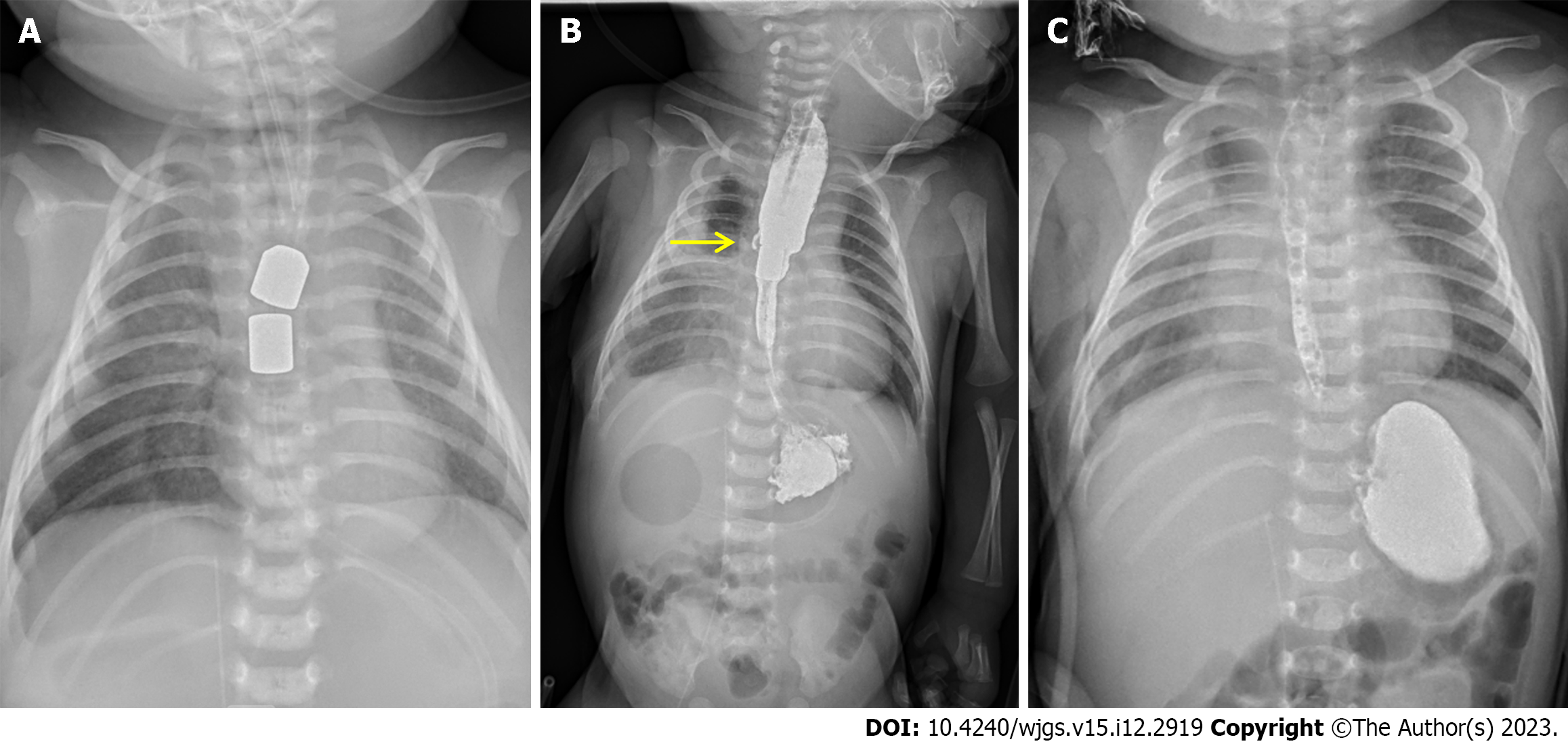
Figure 4 Postoperative X-ray films.
A: Plain film on the 1st postoperative day; B: Film on the 15th postoperative day (yellow arrow: The small leakage, contrast agent: iohexol); C: Film after the removal of the magnets (contrast agent: Iohexol).
- Citation: Zhang HK, Li XQ, Song HX, Liu SQ, Wang FH, Wen J, Xiao M, Yang AP, Duan XF, Gao ZZ, Hu KL, Zhang W, Lv Y, Zhou XH, Cao ZJ. Primary repair of esophageal atresia Gross type C via thoracoscopic magnetic compression anastomosis: A case report. World J Gastrointest Surg 2023; 15(12): 2919-2925
- URL: https://www.wjgnet.com/1948-9366/full/v15/i12/2919.htm
- DOI: https://dx.doi.org/10.4240/wjgs.v15.i12.2919


