Copyright
©The Author(s) 2023.
World J Gastrointest Surg. Dec 27, 2023; 15(12): 2799-2808
Published online Dec 27, 2023. doi: 10.4240/wjgs.v15.i12.2799
Published online Dec 27, 2023. doi: 10.4240/wjgs.v15.i12.2799
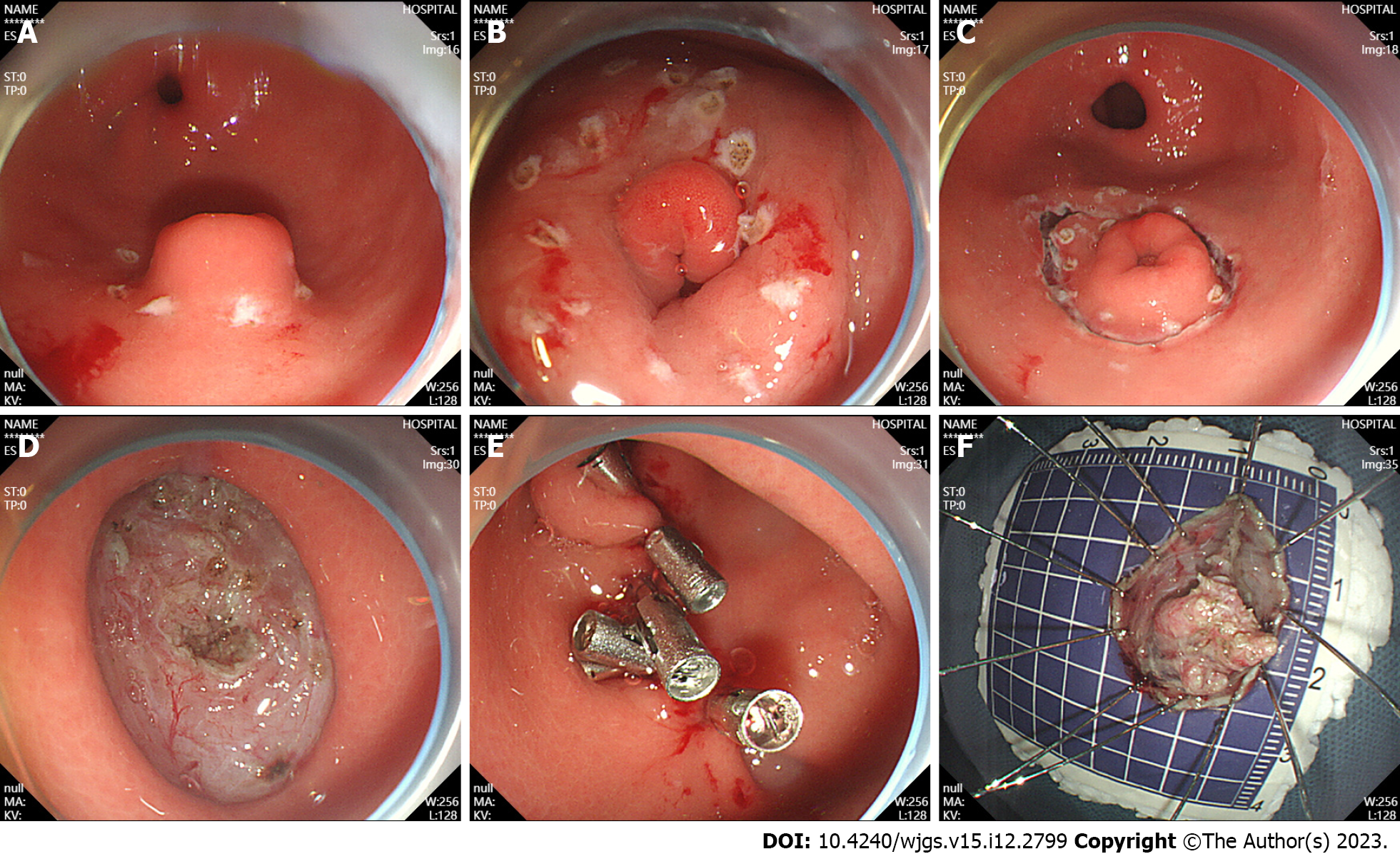
Figure 1 Endoscopic submucosal dissection.
A: Ectopic pancreas was located in the gastric antrum; B: Marked incision line; C: The surrounding tissues of the tumor along the marker were cut; D: Complete tumor resection; E: Intraoperative precautions. F: Removal of the lesions.
Figure 2 Laparoscopic resection.
A: Laparoscopic view of tumor resection using laparoscopic linear anastomosis; B: Laparoscopic view of wedge resection after incision of the gastric wall to exsanguinate the tumor; C: Suture and reinforcement of the remnant gastric wall; D: Pathological specimen.
Figure 3 Pathological view of a gastric ectopic pancreas.
A: Pancreatic tissue (black arrow) and gastric mucosal tissue (white arrow), (magnification, × 40); B: Numerous acini (orange arrow) were observed in pancreatic tissue (magnification, × 100); C: Ducts were observed in the pancreatic tissue (white arrow) (magnification, × 100); D: Islet cells were observed in the pancreatic tissue (orange arrow) (magnification, × 200).
Figure 4 Computed tomography images and endoscopic ultrasound images.
A: The arterial phase of enhanced computed tomography showed thickening of the gastric antrum (orange arrow); B: Lesion morphology on endoscopic ultrasound (white arrow).
- Citation: Zheng HD, Huang QY, Hu YH, Ye K, Xu JH. Laparoscopic resection and endoscopic submucosal dissection for treating gastric ectopic pancreas. World J Gastrointest Surg 2023; 15(12): 2799-2808
- URL: https://www.wjgnet.com/1948-9366/full/v15/i12/2799.htm
- DOI: https://dx.doi.org/10.4240/wjgs.v15.i12.2799


