Copyright
©The Author(s) 2023.
World J Gastrointest Surg. Nov 27, 2023; 15(11): 2657-2662
Published online Nov 27, 2023. doi: 10.4240/wjgs.v15.i11.2657
Published online Nov 27, 2023. doi: 10.4240/wjgs.v15.i11.2657
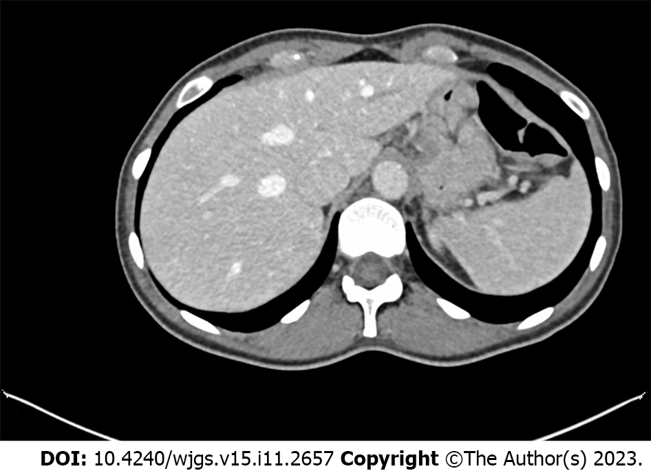
Figure 1 Nodulary injury on the anterior surface of the fundus/gastric body.
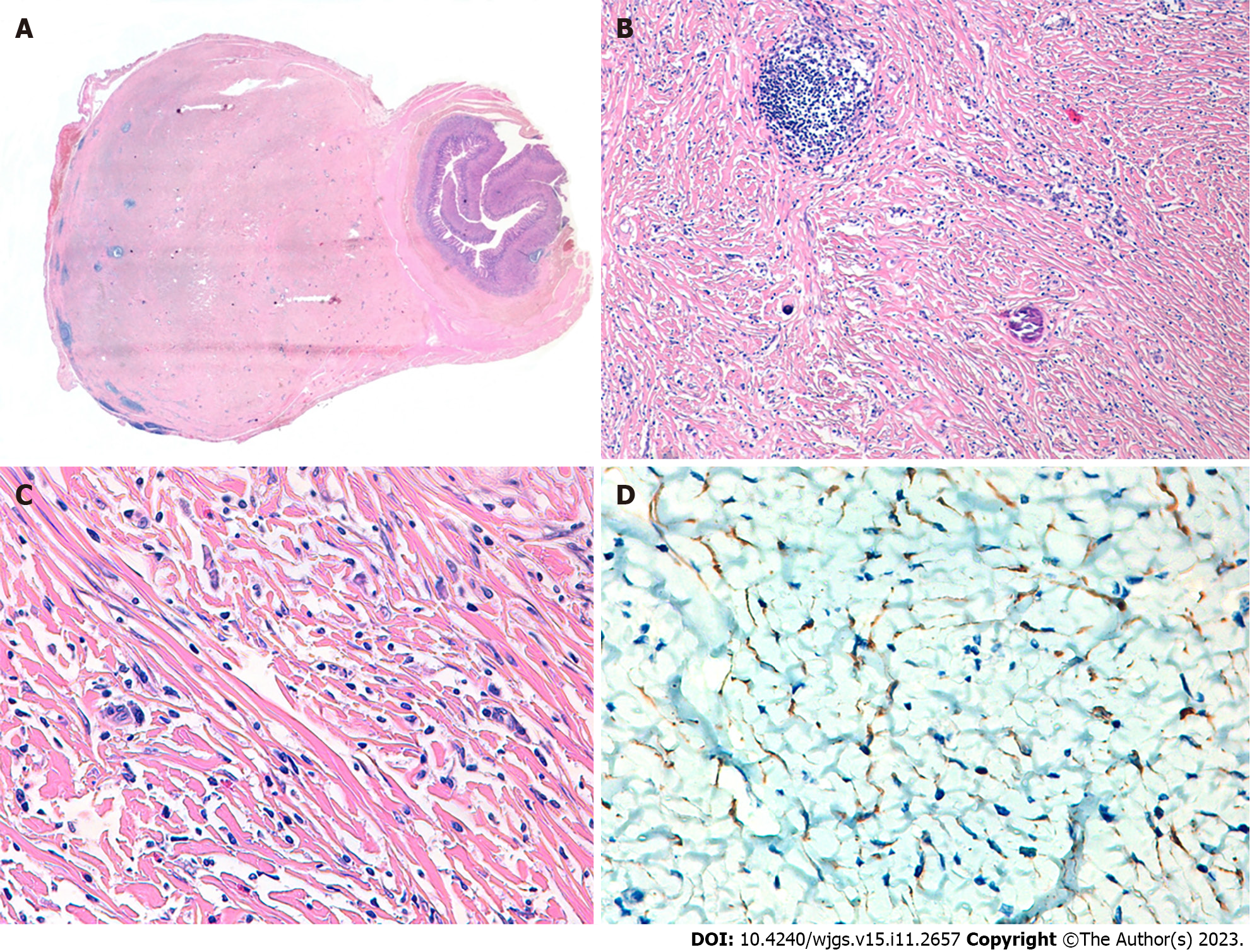
Figure 2 Histopathological analysis and immunohistochemical examination of the resected specimen.
A: Panoramic image of the lesion; B: Mesenquimal injury and psamomatous calcifications; C: Mesenquimal lesion with infiltrate of eosinophils, plasma cells and masts cells; D: Positive expression of Cytokeratins AE1-AE3.
- Citation: Fernandez Rodriguez M, Artuñedo Pe PJ, Callejas Diaz A, Silvestre Egea G, Grillo Marín C, Iglesias Garcia E, Lucena de La Poza JL. Gastric inflammatory myofibroblastic tumor, a rare mesenchymal neoplasm: A case report. World J Gastrointest Surg 2023; 15(11): 2657-2662
- URL: https://www.wjgnet.com/1948-9366/full/v15/i11/2657.htm
- DOI: https://dx.doi.org/10.4240/wjgs.v15.i11.2657


