Copyright
©The Author(s) 2023.
World J Gastrointest Surg. Nov 27, 2023; 15(11): 2639-2645
Published online Nov 27, 2023. doi: 10.4240/wjgs.v15.i11.2639
Published online Nov 27, 2023. doi: 10.4240/wjgs.v15.i11.2639
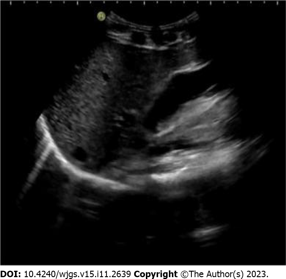
Figure 1
ltrasound examination suggested the lamellar middle and low echo in the gallbladder cavity.
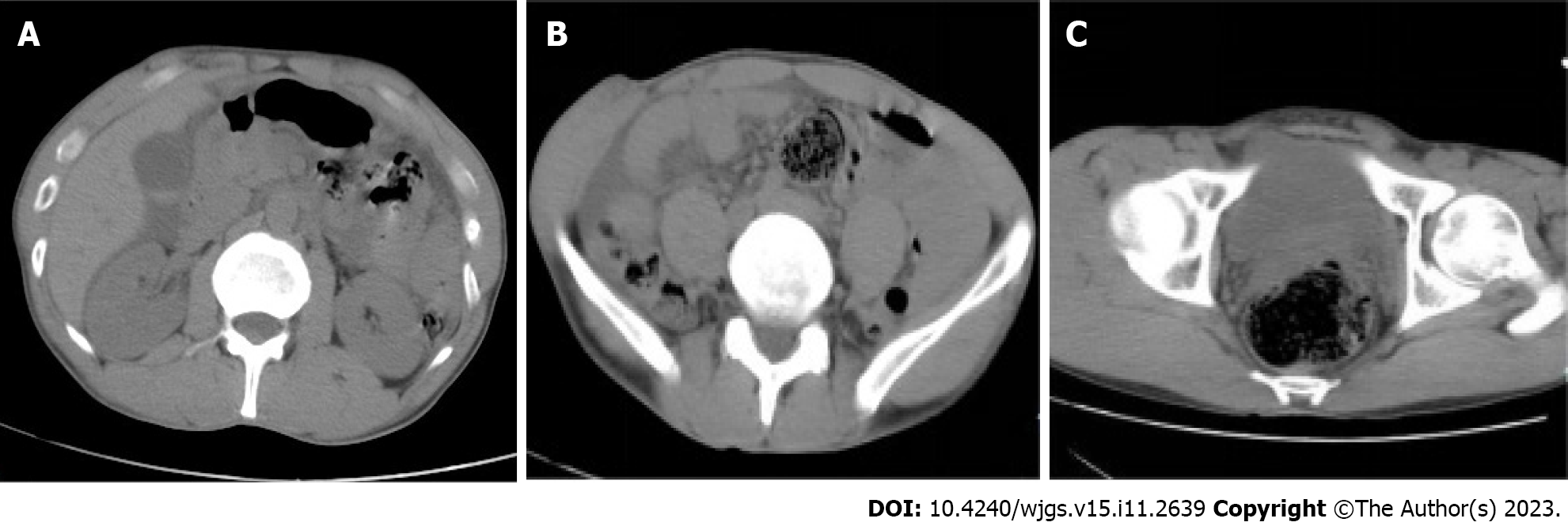
Figure 2 Abdominal computed tomography examination results.
A: This computed tomography (CT) image showed a little high-density area in the gallbladder and cholestasis; B: This CT image indicated abdominal dropsy; C: This CT image showed pelvic effusion, rectal dilatation and fecal accumulation.
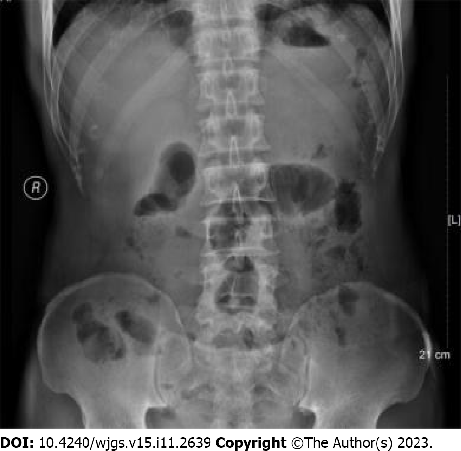
Figure 3
Abdominal anteroposterior radiograph showed that part of the intestine in the middle abdomen is dilated.
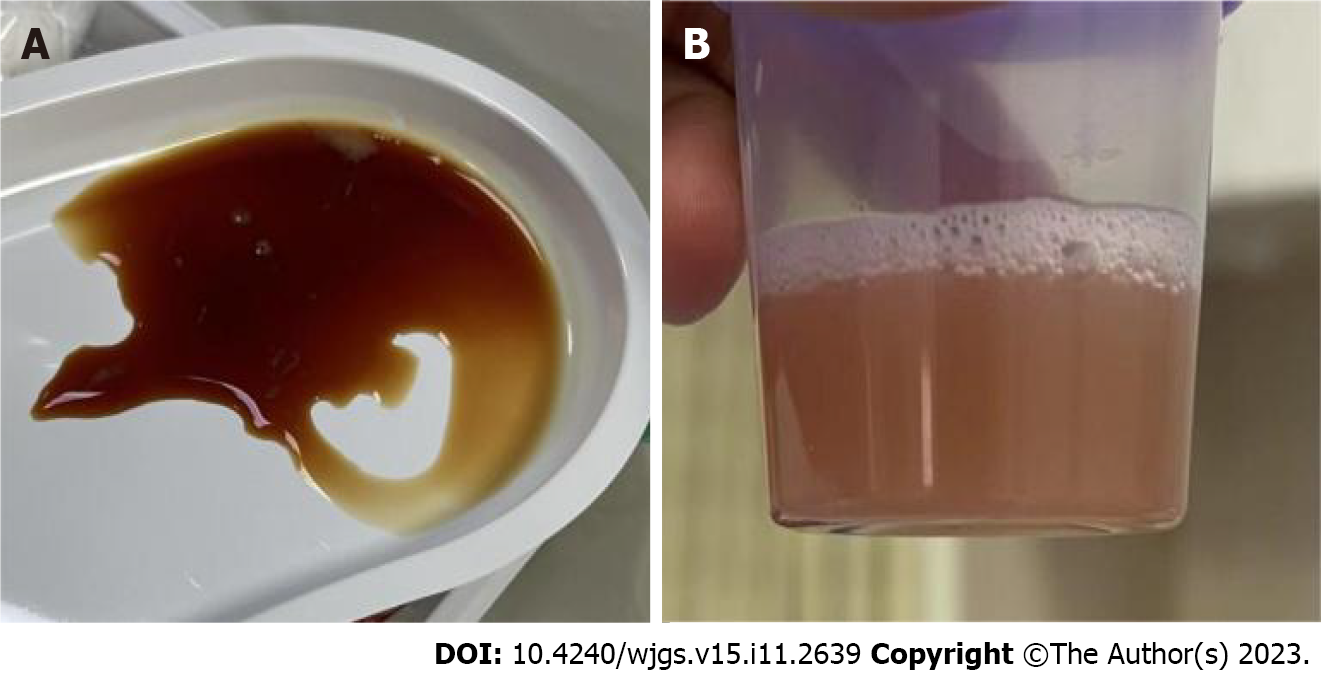
Figure 4 Results of diagnostic abdominal puncture.
A: Light red liquid through the abdominal hole was extracted; B: The puncture drainage fluid was sent to the laboratory for Amylase determination.
Figure 5 The second computed tomography examination results and intraoperative images.
A: The computed tomography (CT) image indicated that abdominal and pelvic effusion increased; B: The abdominal CT showed cholestasis and a slightly high-density gallbladder; C: This image showed that a lacerated wound on the anterior wall of the gallbladder, about 4 cm × 1 cm.
- Citation: Liu DL, Pan JY, Huang TC, Li CZ, Feng WD, Wang GX. Isolated traumatic gallbladder injury: A case report. World J Gastrointest Surg 2023; 15(11): 2639-2645
- URL: https://www.wjgnet.com/1948-9366/full/v15/i11/2639.htm
- DOI: https://dx.doi.org/10.4240/wjgs.v15.i11.2639


