Copyright
©The Author(s) 2023.
World J Gastrointest Surg. Nov 27, 2023; 15(11): 2627-2638
Published online Nov 27, 2023. doi: 10.4240/wjgs.v15.i11.2627
Published online Nov 27, 2023. doi: 10.4240/wjgs.v15.i11.2627
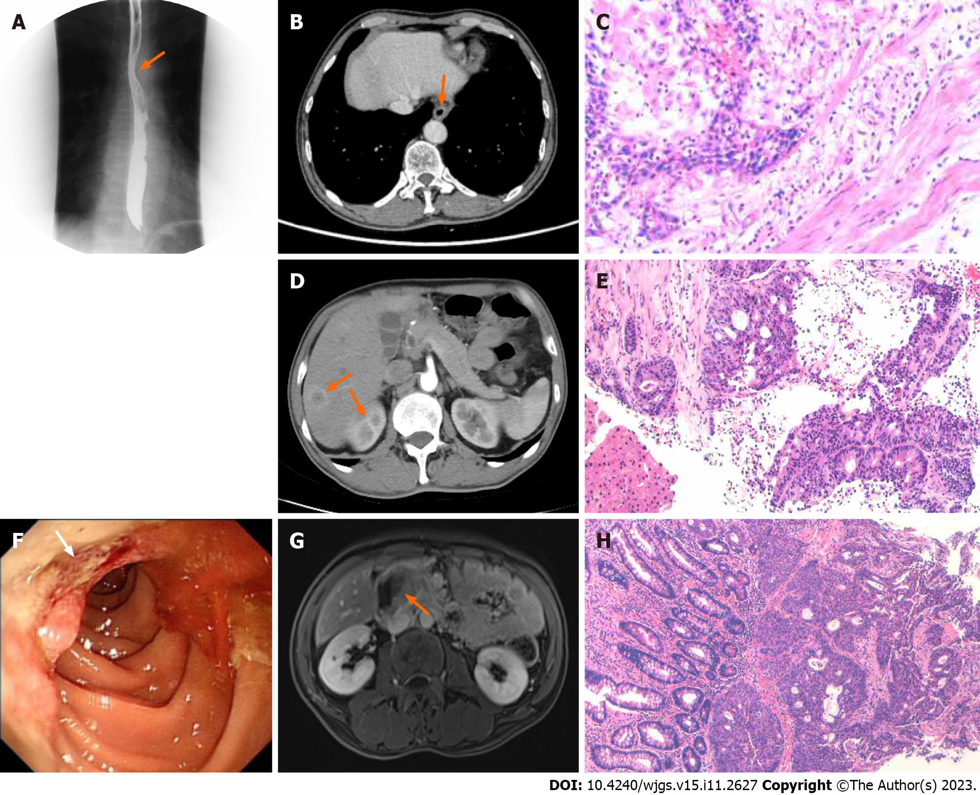
Figure 1 Diagnostic information of primary carcinomas and metastases.
A: The upper gastrointestinal tract barium meal revealed a localization in the lower-middle esophagus on February 20, 2021; B: The enhanced computed tomography (CT) scan of the chest and upper abdomen showed thickness and enhancement of the lower esophagus wall on February 19, 2021; C: Postoperative pathology revealed that the tumor was completely located in the esophagus on February 24, 2021. It was a highly differentiated squamous cell carcinoma (original magnification × 200); D: Abdominal CT enhancement showed multiple metastatic nodules in the liver on September 18, 2021; E: “A needle biopsy of liver mass” was done under ultrasound guidance, and the pathology suggested that the liver lesion was compatible with invasive intermediate differentiated adenocarcinoma on October 19, 2021 (original magnification × 200); F: The gastroscopy revealed an ulcerated neoplasm in the descending portion of the duodenum on October 13, 2021; G: Abdominal magnetic resonance imaging showed an occupancy in the descending and horizontal parts of the duodenum on October 10, 2021; H: Pathology of needle biopsy revealed a medium-low differentiated adenocarcinoma (original magnification × 100).
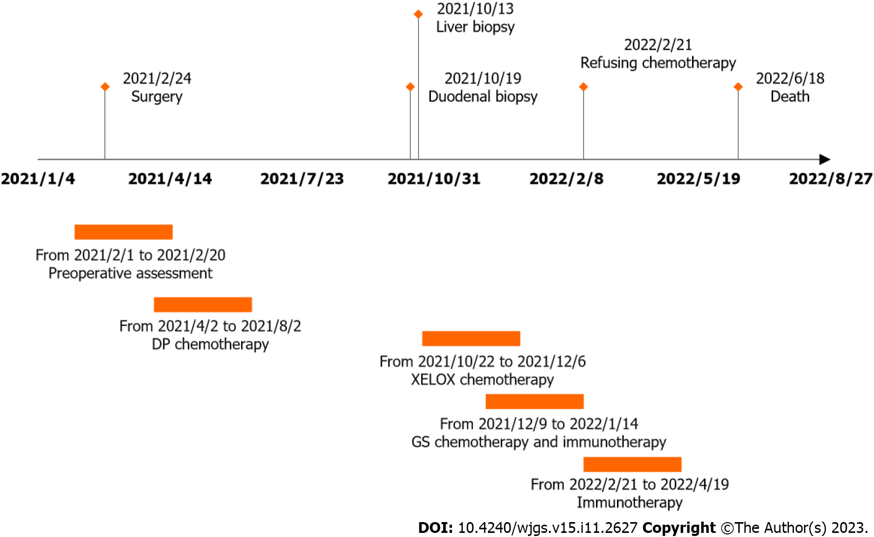
Figure 2 Time points correspond to the diagnostic and therapeutic process.
DP: Doxorubicin and platinum; GS: Gemcitabine and s-1.
- Citation: Huang CC, Ying LQ, Chen YP, Ji M, Zhang L, Liu L. Metachronous primary esophageal squamous cell carcinoma and duodenal adenocarcinoma: A case report and review of literature. World J Gastrointest Surg 2023; 15(11): 2627-2638
- URL: https://www.wjgnet.com/1948-9366/full/v15/i11/2627.htm
- DOI: https://dx.doi.org/10.4240/wjgs.v15.i11.2627


