Copyright
©The Author(s) 2023.
World J Gastrointest Surg. Oct 27, 2023; 15(10): 2362-2366
Published online Oct 27, 2023. doi: 10.4240/wjgs.v15.i10.2362
Published online Oct 27, 2023. doi: 10.4240/wjgs.v15.i10.2362
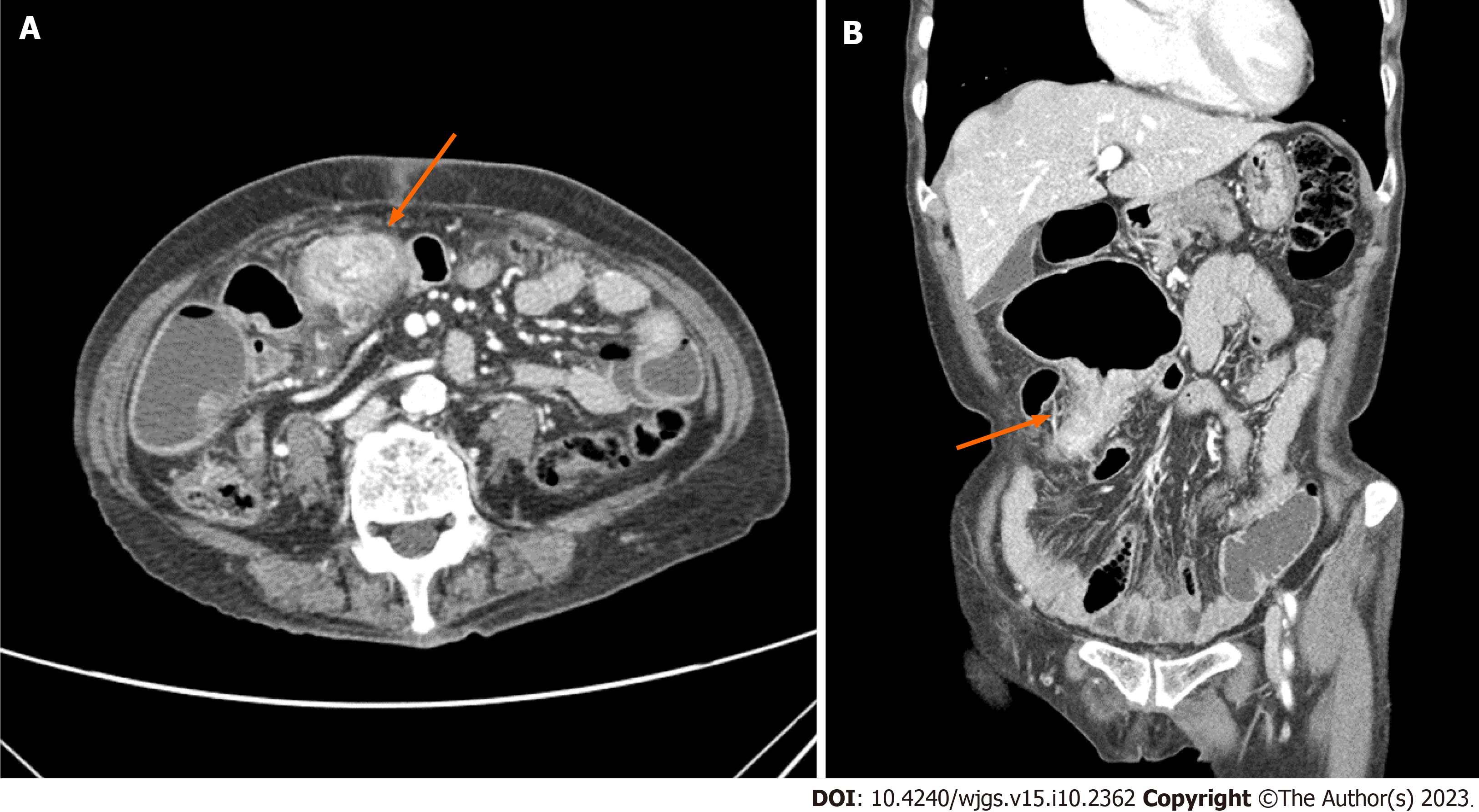
Figure 1 Computed tomography scan demonstrating irregular thickening of the distal ileum, resulting in proximal small bowel dilatation (arrow).
A: Axial view; B: Coronal view.
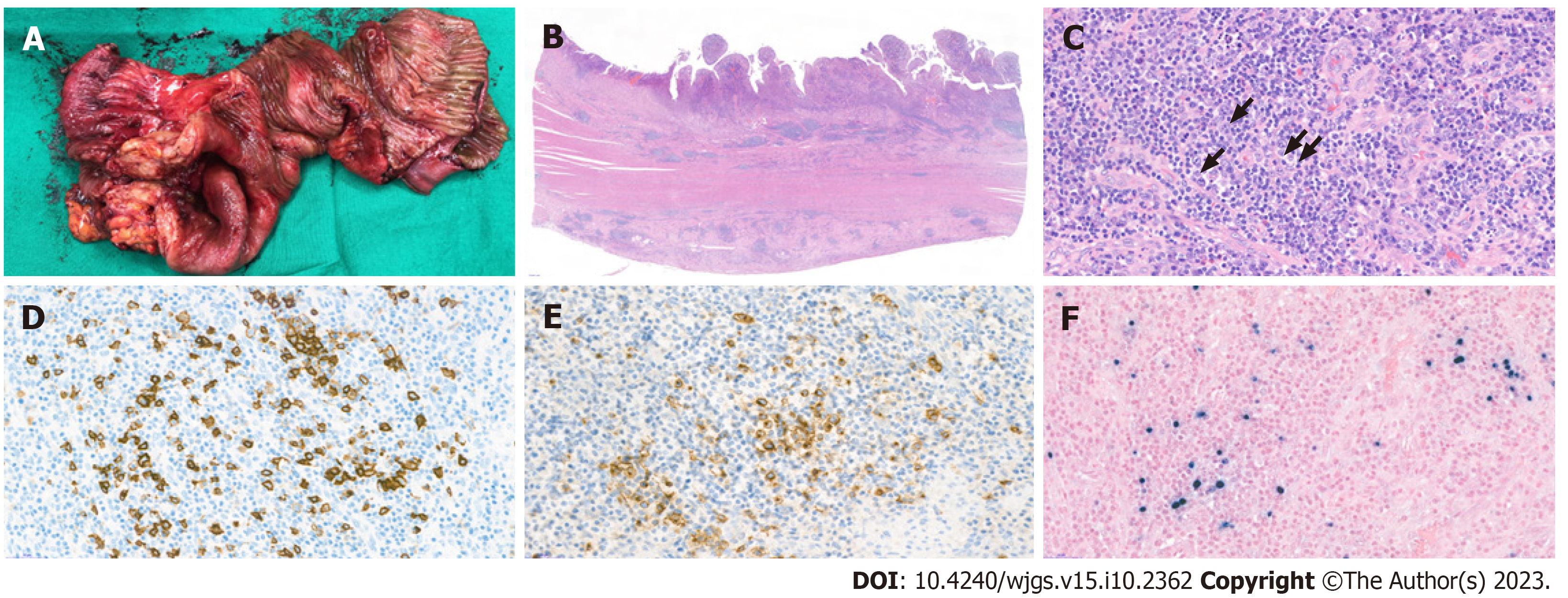
Figure 2 Histopathological analysis of the resected specimen.
A: The resected specimen showed a single ulcerative lesion with luminal obstruction; B: Histopathologically, the mucosal surface was ulcerated with granulation tissue formation. Beneath the ulcer, the specimen revealed marked infiltration of various inflammatory cells as well as dense fibrosis in all layers of the colon wall (hematoxylin–eosin stain, scan view); C: The infiltrated inflammatory cells consisted of lymphocytes, plasma cells, eosinophils and neutrophils, as well as a few scattered large atypical lymphoid cells (arrow) (hematoxylin–eosin stain, original magnification, 400×); D–F: The large atypical lymphoid cells were CD20-positive, CD30-positive, and Epstein–Barr virus (EBV)-positive. CD20 (D), CD30 (E), and in situ hybridization for EBV-encoded RNA (F) (original magnification, 400×).
- Citation: Song JH, Choi JE, Kim JS. Mucocutaneous ulcer positive for Epstein–Barr virus, misdiagnosed as a small bowel adenocarcinoma: A case report. World J Gastrointest Surg 2023; 15(10): 2362-2366
- URL: https://www.wjgnet.com/1948-9366/full/v15/i10/2362.htm
- DOI: https://dx.doi.org/10.4240/wjgs.v15.i10.2362


