Copyright
©The Author(s) 2023.
World J Gastrointest Surg. Oct 27, 2023; 15(10): 2351-2356
Published online Oct 27, 2023. doi: 10.4240/wjgs.v15.i10.2351
Published online Oct 27, 2023. doi: 10.4240/wjgs.v15.i10.2351
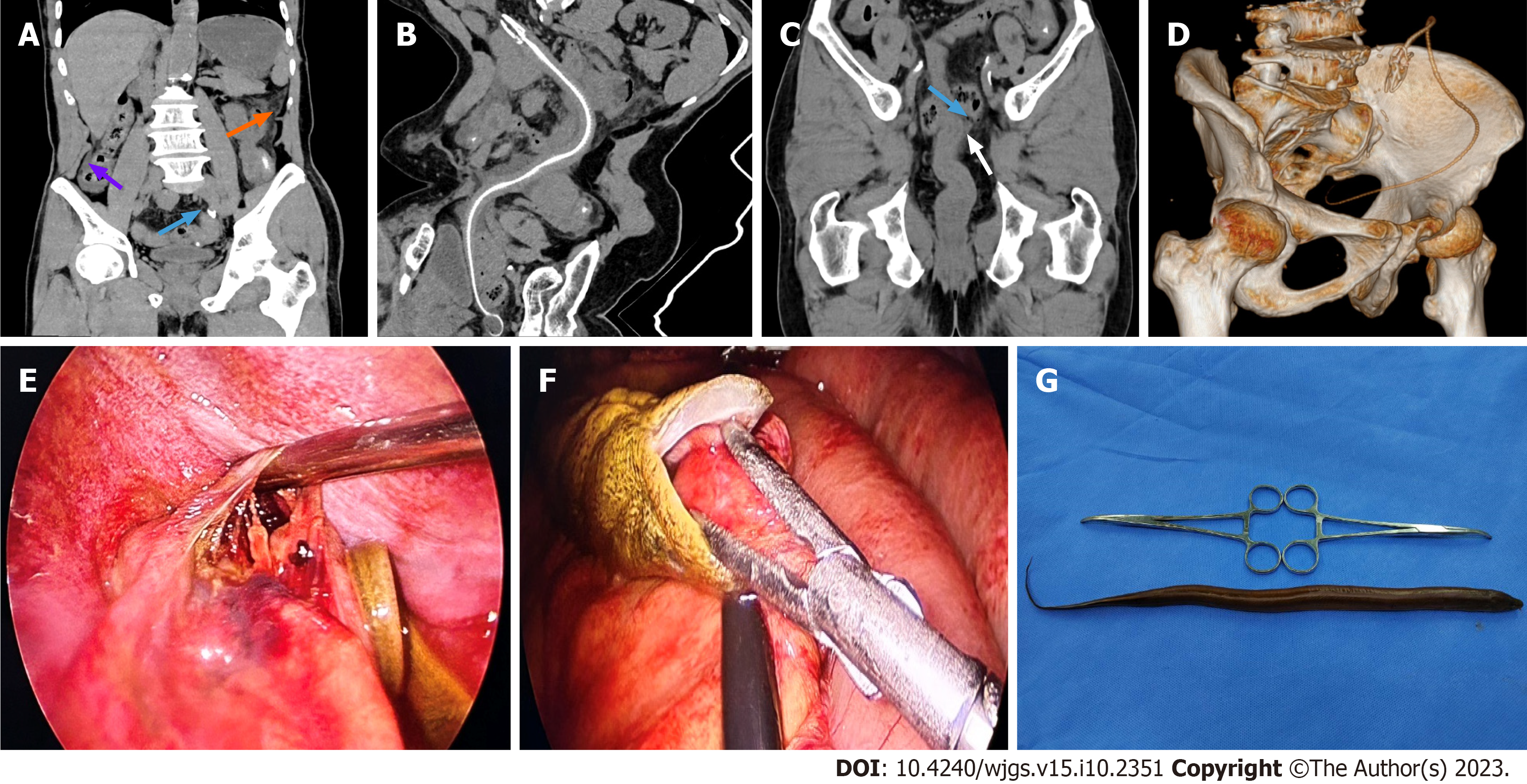
Figure 1 Imaging and laparoscopic exploration.
A: Computed tomography (CT) with multi-plane reconstruction revealed scattered exudation, peritoneal thickening (orange arrow), peritoneal effusion (purple arrow), and free gas (blue arrow) in the abdominal cavity, suggesting gastrointestinal perforation complicated with acute peritonitis; B: Curved planar reconstruction (CPR) of CT images showed an abdominal Monopterus albus that has bitten the mesentery, indicating a foreign body outside the intestinal cavity; C: CPR of CT images revealed rough and raised outer margin of the wall of the mid-rectum (white arrow), as well as exudation and free gas in the surrounding mesentery (blue arrow), indicating a perforation in the mid-rectum; D: Volume reconstruction revealed clear and complete Monopterus albus bone morphology in the abdominal and pelvic cavities; E: Laparoscopic exploration revealed a large perforation in the mid-rectum, with a diameter approximating 1.5 cm; F: Laparoscopic exploration showed the Monopterus albus has perforated the mesentery of the small intestine; G: During the operation, the dead Monopterus albus was taken out, with a length of about 40 cm.
- Citation: Yang JH, Lan JY, Lin AY, Huang WB, Liao JY. Three-dimensional computed tomography reconstruction diagnosed digestive tract perforation and acute peritonitis caused by Monopterus albus: A case report. World J Gastrointest Surg 2023; 15(10): 2351-2356
- URL: https://www.wjgnet.com/1948-9366/full/v15/i10/2351.htm
- DOI: https://dx.doi.org/10.4240/wjgs.v15.i10.2351


