Copyright
©The Author(s) 2023.
World J Gastrointest Surg. Oct 27, 2023; 15(10): 2343-2350
Published online Oct 27, 2023. doi: 10.4240/wjgs.v15.i10.2343
Published online Oct 27, 2023. doi: 10.4240/wjgs.v15.i10.2343
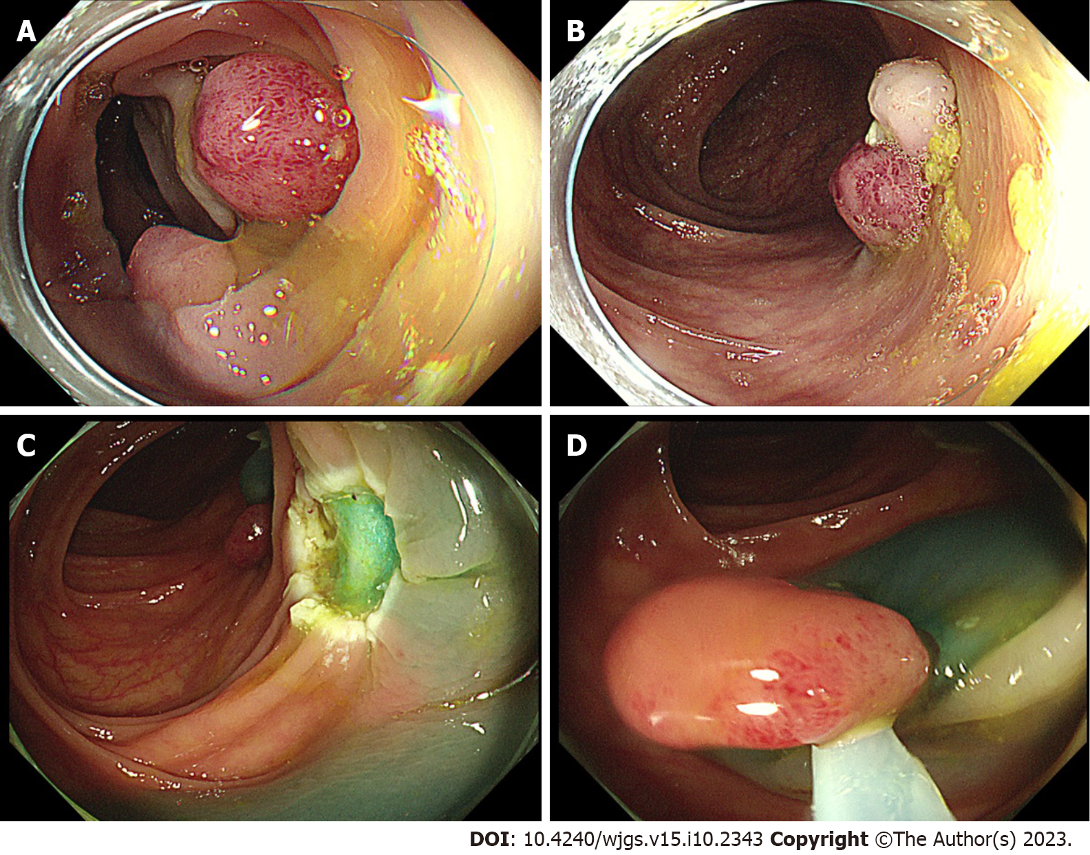
Figure 1 Clinical findings during colonoscopy.
A and B: The first colonoscopy (day 3) showed multiple broad-based or pedicled polyps with diameters ranging from 0.5 to 2.0 cm in the cecum, ascending colon, transverse colon, descending colon, and sigmoid colon; C: The second colonoscopy (day 9) showed multiple ulcerations in the transverse colon and descending colon, which were wounds from the first surgery; D: On the second colonoscopy (day 9), three polyps (0.8-1.2 cm) scattered in the descending and sigmoid colons were removed by endoscopic mucosal resection.
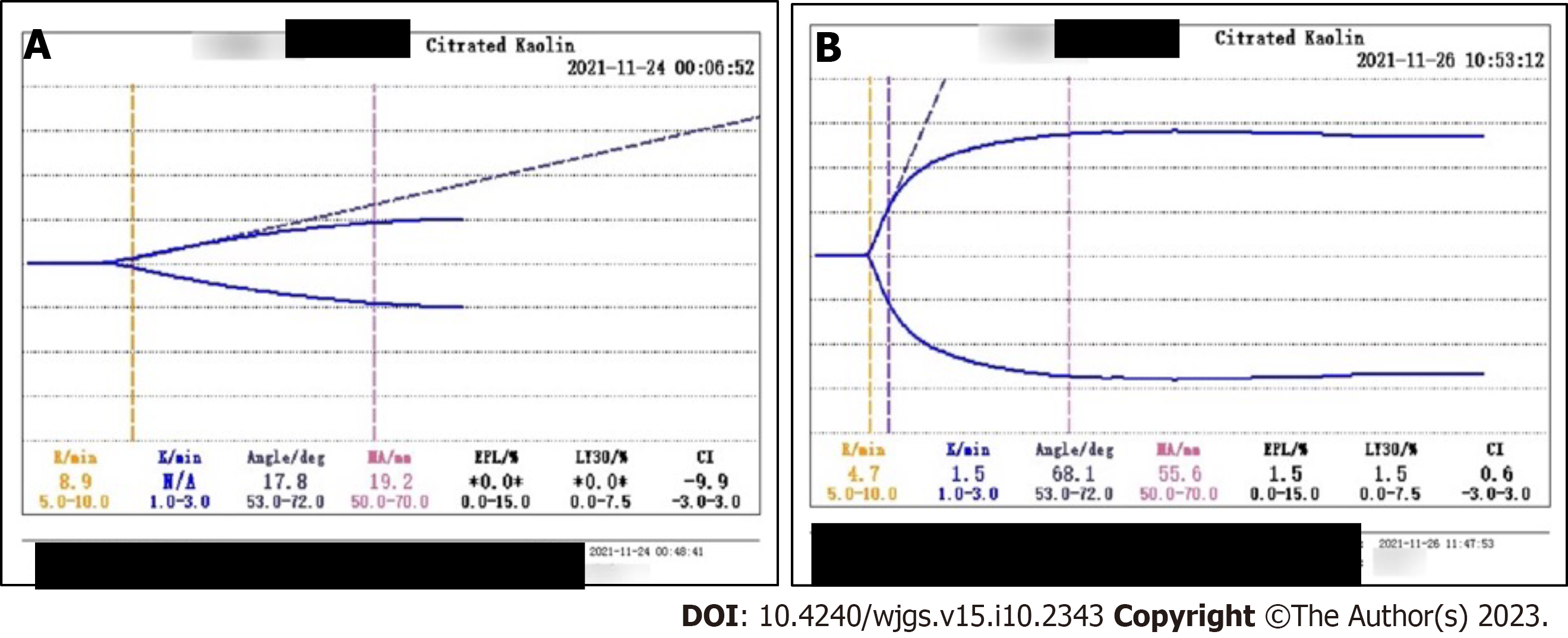
Figure 2 Thrombelastogram.
A: On day 9, the comprehensive coagulation index was -9.9, fibrinogen function α-angle (deg) was 17.8, K-time exceeded the upper limit, and maximum amplitude representing platelet function was 19.2 mm, showing severe coagulation dysfunction; B: On day 9, the indicators returned to normal.
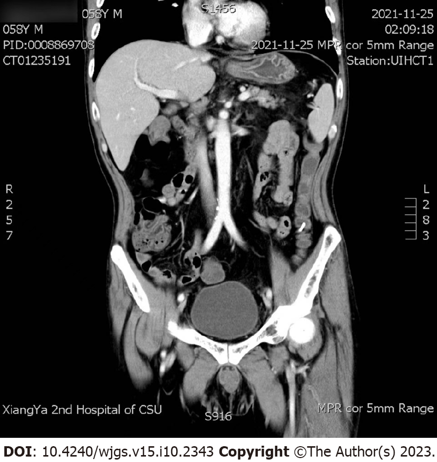
Figure 3 Abdominal computed tomography and superior mesenteric artery computed tomography angiography.
It showed no obvious intestinal perforation or lumbar lesion.
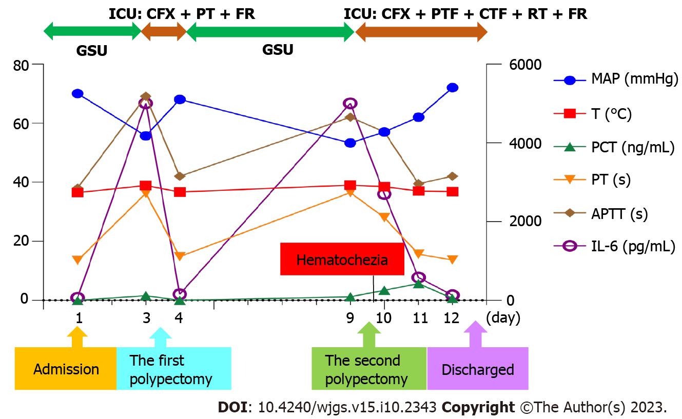
Figure 4 Clinical course with laboratory results and treatment.
GSU: Gastrointestinal surgery unit; ICU: Intensive care unit; CFX: Cefoxitin; PTF: Plasma transfusion; FR: Fluid resuscitation; CTF: Coprecipitation transfusion; RT: Red cells transfusion; MAP: Mean arterial pressure; T: Daily maximum body temperature; PCT: Procalcitonin; PT: Prothrombin time; APTT: Activated partial thromboplastin time; IL-6: Interleukin 6.
- Citation: Chen FZ, Ouyang L, Zhong XL, Li JX, Zhou YY. Postpolypectomy syndrome without abdominal pain led to sepsis/septic shock and gastrointestinal bleeding: A case report. World J Gastrointest Surg 2023; 15(10): 2343-2350
- URL: https://www.wjgnet.com/1948-9366/full/v15/i10/2343.htm
- DOI: https://dx.doi.org/10.4240/wjgs.v15.i10.2343


