Copyright
©The Author(s) 2023.
World J Gastrointest Surg. Oct 27, 2023; 15(10): 2133-2141
Published online Oct 27, 2023. doi: 10.4240/wjgs.v15.i10.2133
Published online Oct 27, 2023. doi: 10.4240/wjgs.v15.i10.2133
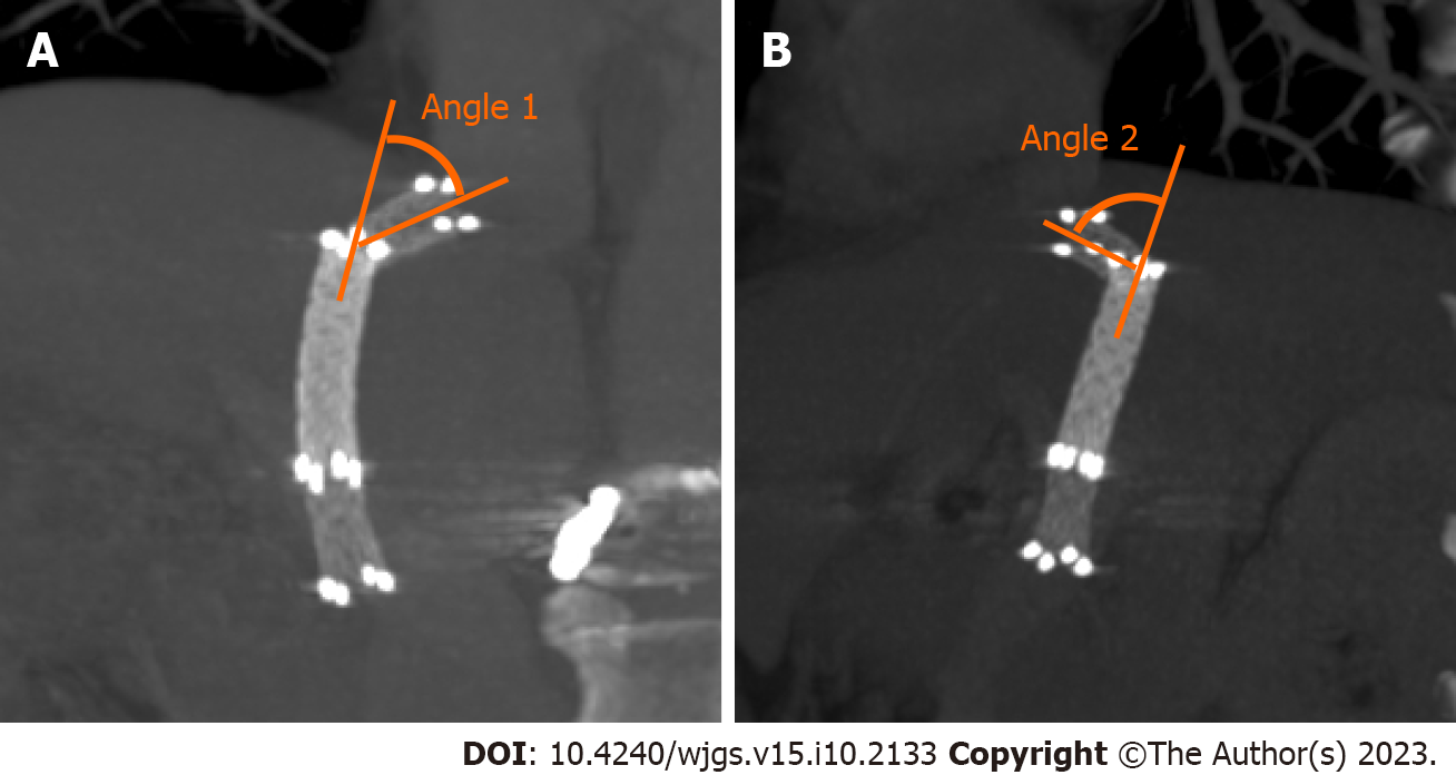
Figure 1 Computed tomography images showing angle 1 and angle 2.
A: The angle between the proximal end of the bare metal stent and the stent-graft is measured angle 1 in the coronal plane; B: Angle 2 in the sagittal plane.
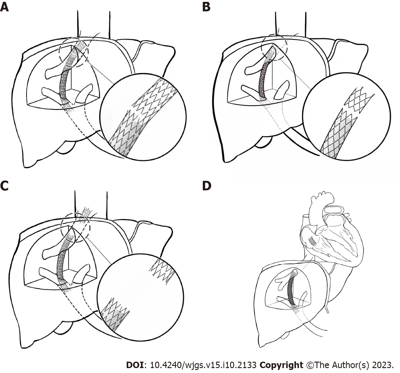
Figure 2 Types of stent fracture after the transjugular intrahepatic portosystemic shunt procedure.
A: Type I partial fractures of the stent struts; B: Type II annular fractures of the stent struts, but without displacement; C and D: Type III stent transection with fractured stent displacement (C, IIIa: Small displacement; D, IIIb: Displacement of the stents from the original vessels into adjacent structures).
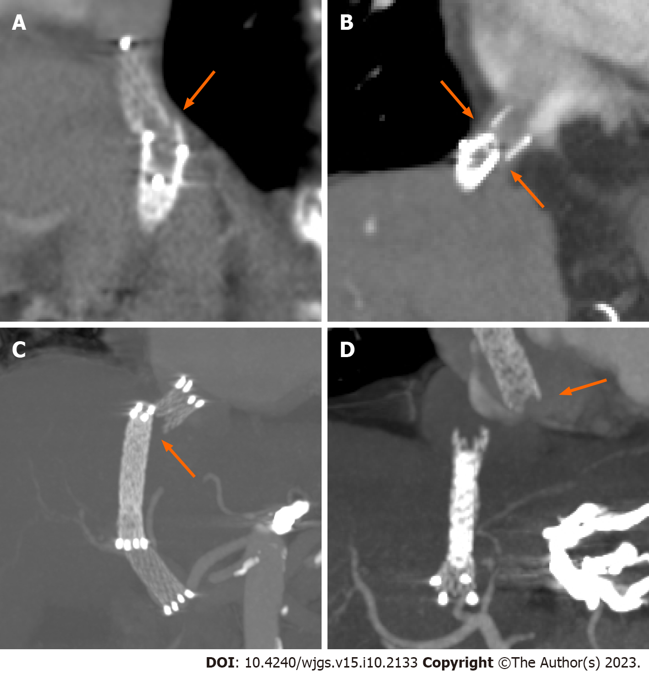
Figure 3 Representative computed tomography images of the types of stent fracture after transjugular intrahepatic portosystemic shunt placement.
The orange arrows show the position and displacement of stent fractures. A: Type I partial fracture of the stent struts; B: Type II annular fracture of the stent struts; C: Type IIIa fractured stent with minimal displacement and D: Type IIIb fractured stent displaced into the heart chamber.
- Citation: Liu QJ, Cao XF, Pei Y, Li X, Dong GX, Wang CM. Stent fracture after transjugular intrahepatic portosystemic shunt placement using the bare metal stent/stent-graft combination technique. World J Gastrointest Surg 2023; 15(10): 2133-2141
- URL: https://www.wjgnet.com/1948-9366/full/v15/i10/2133.htm
- DOI: https://dx.doi.org/10.4240/wjgs.v15.i10.2133


