Copyright
©The Author(s) 2023.
World J Gastrointest Surg. Jan 27, 2023; 15(1): 114-120
Published online Jan 27, 2023. doi: 10.4240/wjgs.v15.i1.114
Published online Jan 27, 2023. doi: 10.4240/wjgs.v15.i1.114
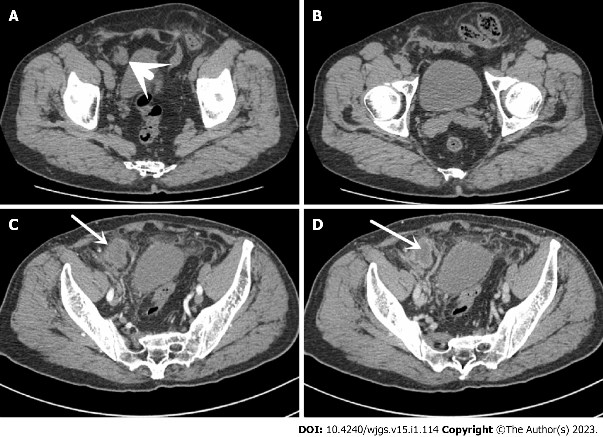
Figure 1 Computed tomography.
A: Abdominal computed tomography (CT) in 2018 reveals a left indirect inguinal hernia, and a hypodense focus (short arrow) in the right lower abdomen; B: The contents of the left indirect inguinal hernia are the sigmoid colon; C: Abdominal CT in 2020 shows a mass on the lateral side of the right umbilical artery (long arrow), with no obvious enhancement in the arterial phase; D: The central part of the mass is found to be more hypointense in the venous phase (long arrow), with circumferential enhancement around the edges of the mass.
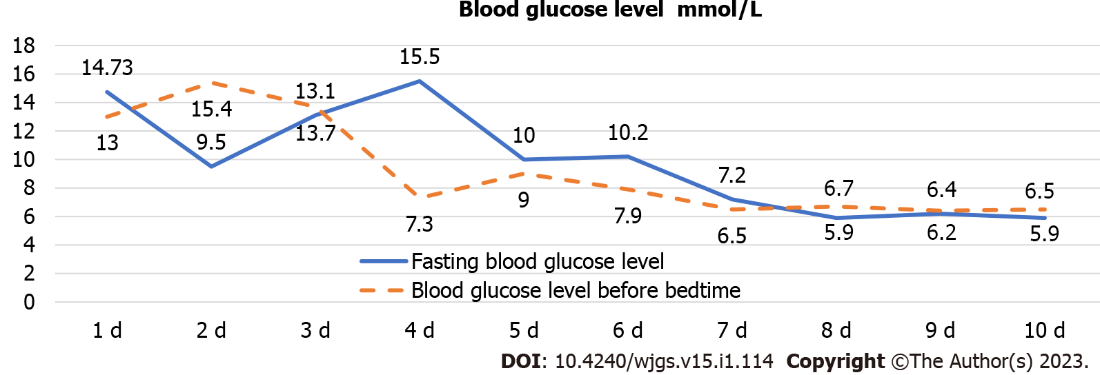
Figure 2
Blood glucose control levels.
Continuous fold is fasting, intermittent line is bedtime.
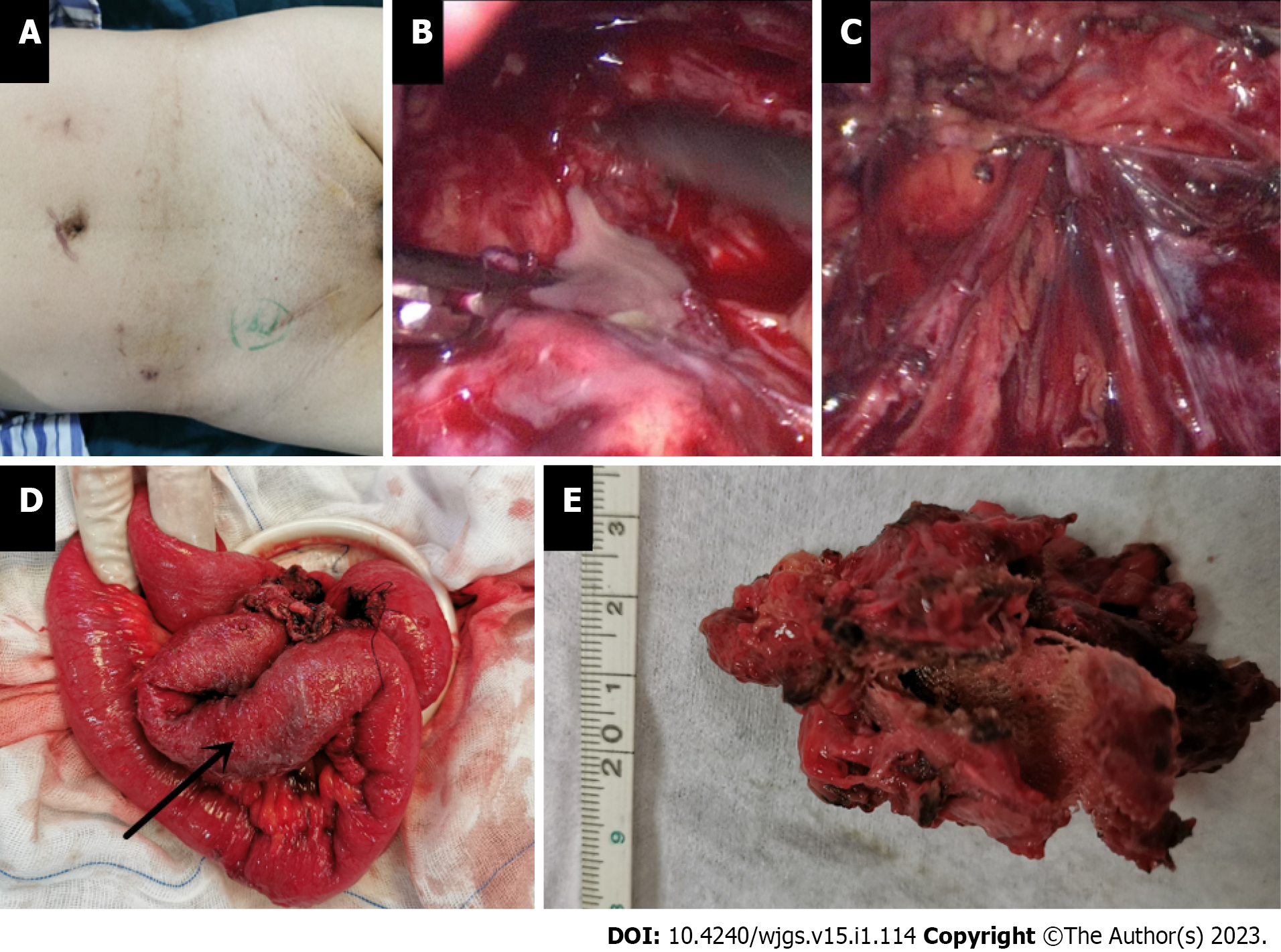
Figure 3 Intraoperative findings.
A: Surgical scar after multiple inguinal hernia repairs; B: Greyish white, purulent, viscous fluid from the mass found intraoperatively; C: Examination of the vas deferens and spermatic vessels after debridement of the mass with no damage and no defective weak areas in the internal ring opening or abdominal wall; D: Partial exfoliation of the ileal canal pulpy muscle layer (arrow) with tortuous intestinal ducts in a mass, closely related to the meshoma; E: Postoperative autopsy reveals a central non-resorbable mesh structure and a cavity in the mass.
- Citation: Wu JF, Chen J, Hong F. Intestinal erosion caused by meshoma displacement: A case report. World J Gastrointest Surg 2023; 15(1): 114-120
- URL: https://www.wjgnet.com/1948-9366/full/v15/i1/114.htm
- DOI: https://dx.doi.org/10.4240/wjgs.v15.i1.114


