Copyright
©The Author(s) 2022.
World J Gastrointest Surg. Jul 27, 2022; 14(7): 714-719
Published online Jul 27, 2022. doi: 10.4240/wjgs.v14.i7.714
Published online Jul 27, 2022. doi: 10.4240/wjgs.v14.i7.714
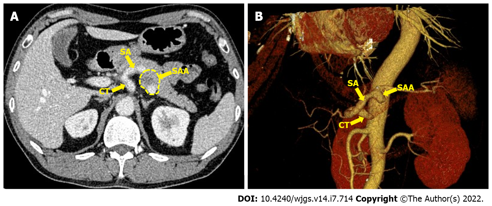
Figure 1 Contrast-enhanced celiac trunk and 3D reconstruction imaging.
A: A 3.5 cm splenic artery aneurysm (SAA) in the proximal splenic artery located in the posterior pancreas; B: 3D reconstruction imaging shows a 3.5 cm SAA at the same location. CT: Celiac trunk; SA: Splenic artery; SAA: Splenic artery aneurysm;
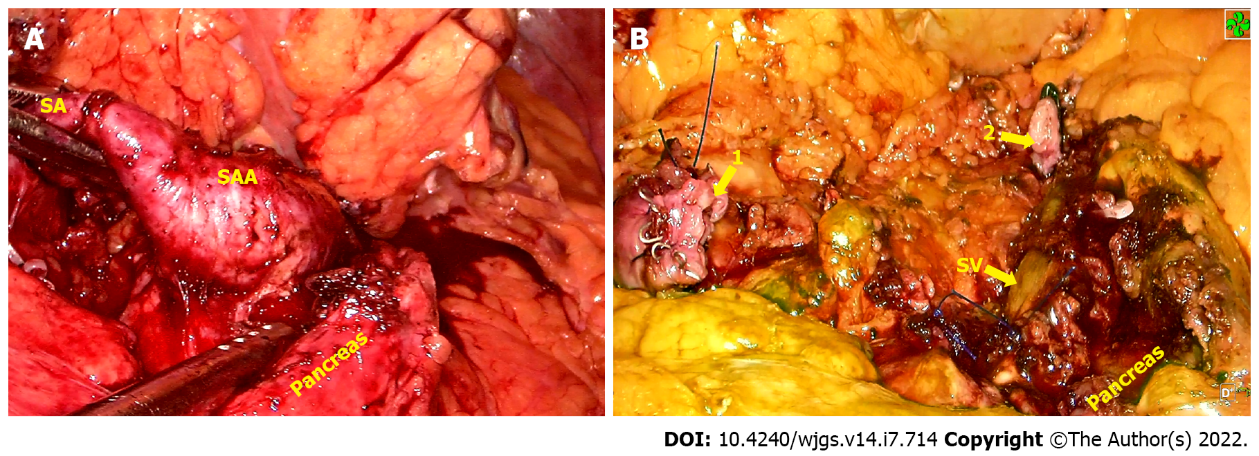
Figure 2 Intraoperative imaging.
A: The splenic artery aneurysm protruded into the pancreatic parenchyma adhered to the surrounding tissues; B: Both the proximal (1) and distal (2) aneurysms were occluded with aneurysmectomy. SA: Splenic artery; SAA: Splenic artery aneurysm; SV: Splenic vein.
Figure 3 Indocyanine green fluorescence imaging at the end of surgery.
A: Spleen before indocyanine green (ICG) injection; B: The whole spleen was stained green 6 min 50 s after ICG injection.
- Citation: Cheng J, Sun LY, Liu J, Zhang CW. Indocyanine green fluorescence imaging for spleen preservation in laparoscopic splenic artery aneurysm resection: A case report. World J Gastrointest Surg 2022; 14(7): 714-719
- URL: https://www.wjgnet.com/1948-9366/full/v14/i7/714.htm
- DOI: https://dx.doi.org/10.4240/wjgs.v14.i7.714


