Copyright
©The Author(s) 2022.
World J Gastrointest Surg. Jun 27, 2022; 14(6): 621-625
Published online Jun 27, 2022. doi: 10.4240/wjgs.v14.i6.621
Published online Jun 27, 2022. doi: 10.4240/wjgs.v14.i6.621
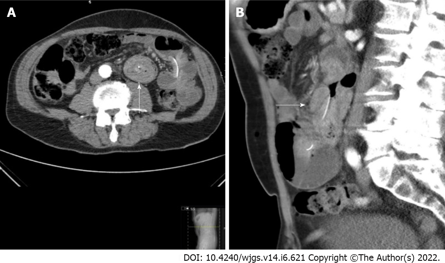
Figure 1 Abdominal computed tomography scan with intravenous contrast in the arterial and portal venous phases of a 73-year-old man with intussusception at the duodenojejunal junction.
A: The transverse section shows a ‘target sign’; B: The sagittal section shows a ‘sausage sign’.
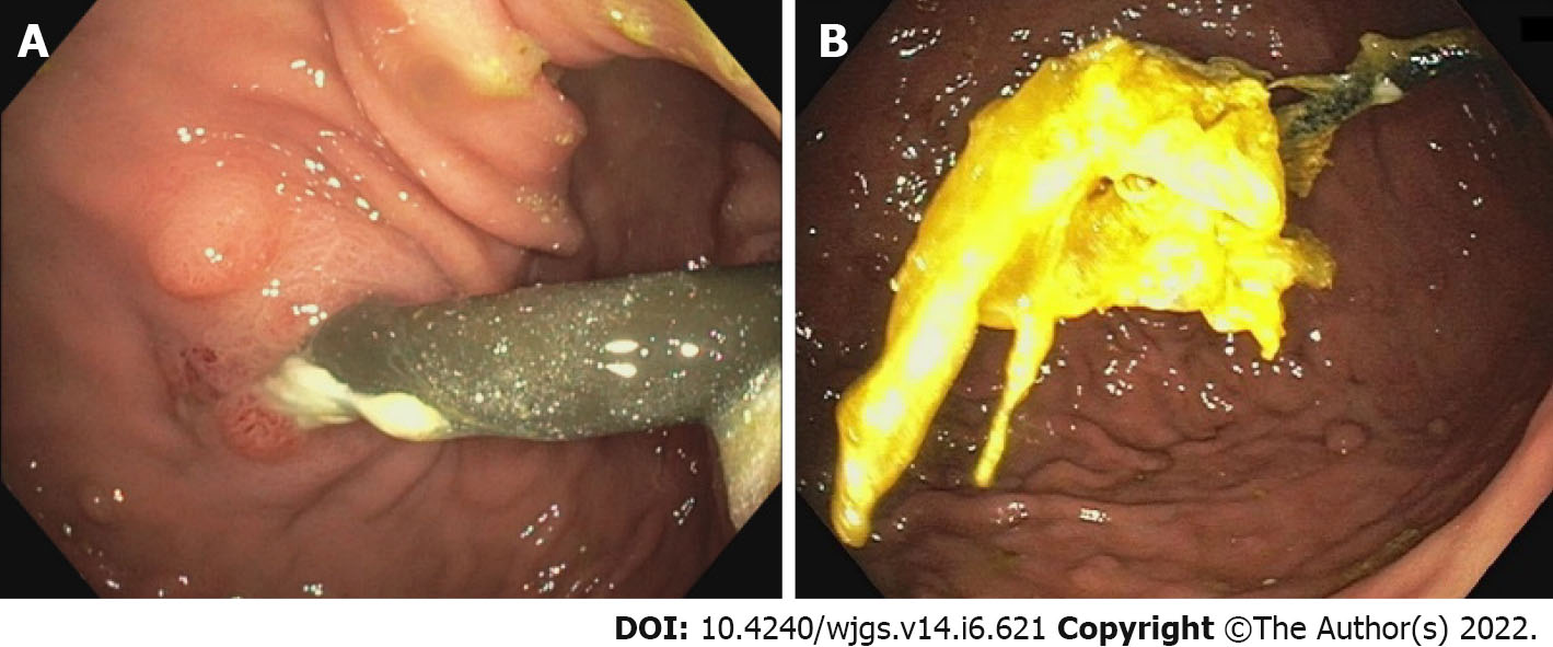
Figure 2 Push enteroscopy: In a 73-year-old man with intussusception.
A: Showing a view of the stomach. Due to traction at the jejunal extension, the button of the percutaneous endoscopic gastrostomy catheter was not situated against the stomach wall; B: Showing the luxated jejunum extension with remnant bezoar after endoscopic reduction.
- Citation: Winters MW, Kramer S, Mazairac AH, Jutte EH, van Putten PG. Bowel intussusception caused by a percutaneously placed endoscopic gastrojejunostomy catheter: A case report. World J Gastrointest Surg 2022; 14(6): 621-625
- URL: https://www.wjgnet.com/1948-9366/full/v14/i6/621.htm
- DOI: https://dx.doi.org/10.4240/wjgs.v14.i6.621


