Copyright
©The Author(s) 2022.
World J Gastrointest Surg. May 27, 2022; 14(5): 506-513
Published online May 27, 2022. doi: 10.4240/wjgs.v14.i5.506
Published online May 27, 2022. doi: 10.4240/wjgs.v14.i5.506
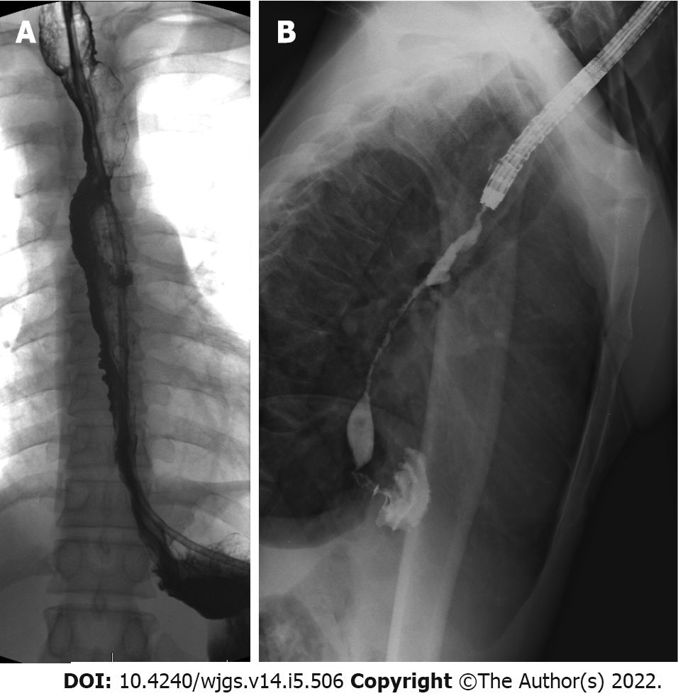
Figure 1 Contrast swallow examination on day nine post injury.
A: Contrast swallow study performed 9 d post injury, already confirming early long-segment stricturing of the oesophagus; B: Fluoroscopic study during endoscopy performed 4 wk post injury, showing high-grade, long-segment oesophageal stricturing.
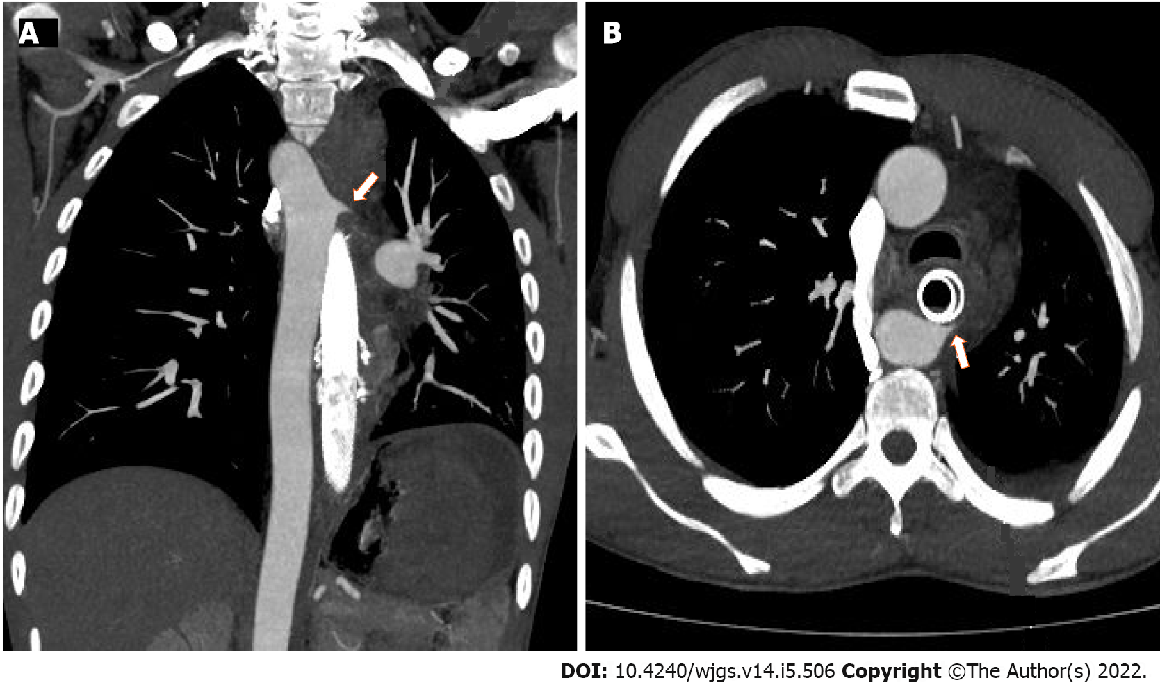
Figure 2 Computed tomography angiogram images confirming the site of the proximal aorto-oesophageal fistula (arrows).
A: Coronal image; B: Axial image.
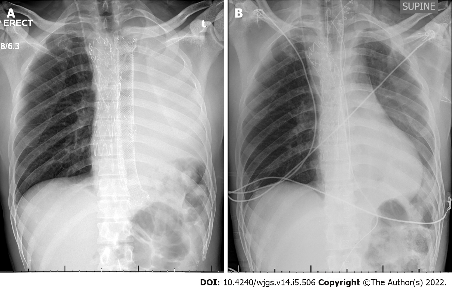
Figure 3 Chest X-ray.
A: Chest X-ray post aortic endovascular repair, showing aortic stent-graft, multiple overlapping stents in the oesophagus and white-out of the left lung, caused by left main bronchus compression by the oesophageal stents; B: Chest X-ray immediately post-operative after retrosternal gastric pull-up and removal of oesophageal stents showing good left lung re-expansion.
- Citation: Scriba MF, Kotze U, Naidoo N, Jonas E, Chinnery GE. Aorto-oesophageal fistula after corrosive ingestion: A case report. World J Gastrointest Surg 2022; 14(5): 506-513
- URL: https://www.wjgnet.com/1948-9366/full/v14/i5/506.htm
- DOI: https://dx.doi.org/10.4240/wjgs.v14.i5.506


