Copyright
©The Author(s) 2022.
World J Gastrointest Surg. Apr 27, 2022; 14(4): 362-369
Published online Apr 27, 2022. doi: 10.4240/wjgs.v14.i4.362
Published online Apr 27, 2022. doi: 10.4240/wjgs.v14.i4.362
Figure 1 Preoperative endoscopy and endoscopic ultrasonography.
A: Upper digestive tract endoscopy showing a submucosal tumor along the greater curvature of the anterior gastric antrum wall; B: Endoscopic ultrasonography showing a mass within the gastric antrum, which originated from the muscularis propria; C: Gastroscopy 3 mo after surgery revealing appropriate incision healing.
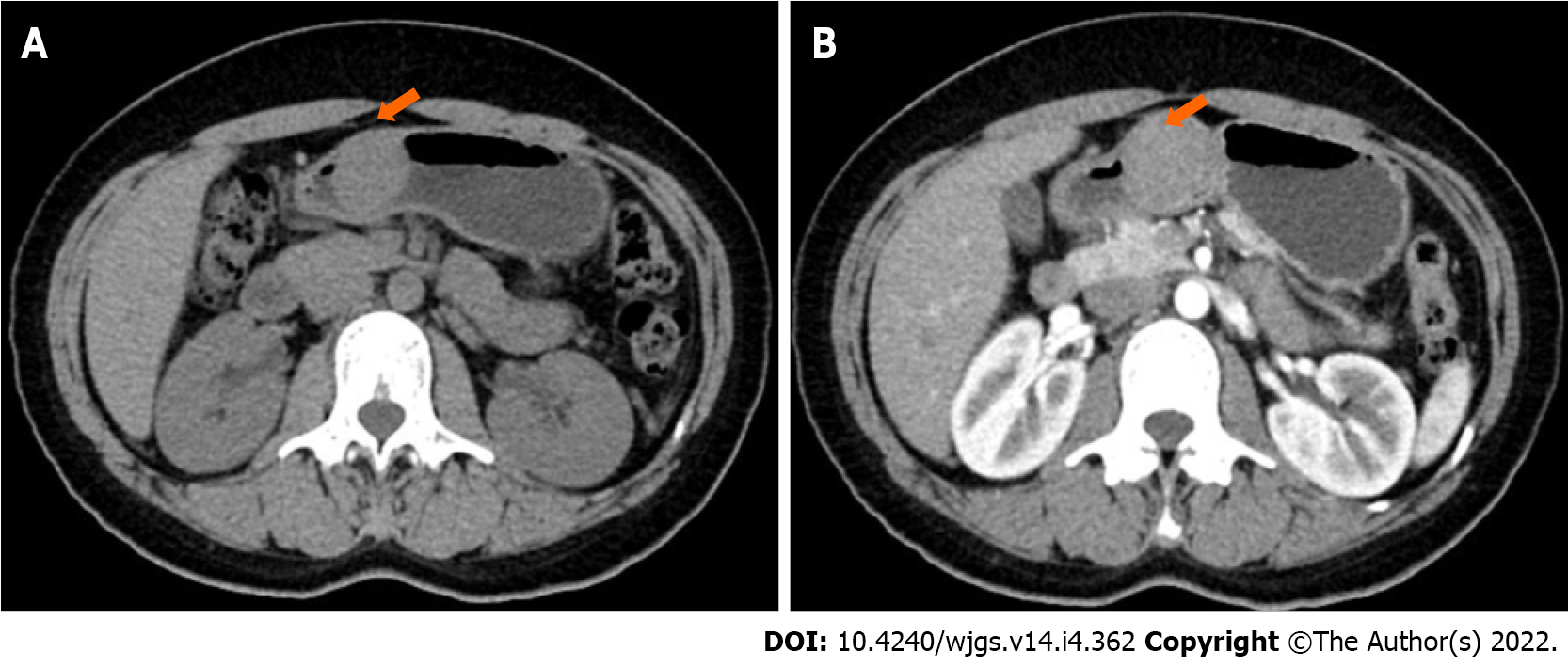
Figure 2 Computed tomography scan.
A: Computed tomography showing an oval mass in the antrum of the stomach, with intracavitary growth; B: Enhanced computed tomography shows obvious enhancement of the mass in the arterial phase.
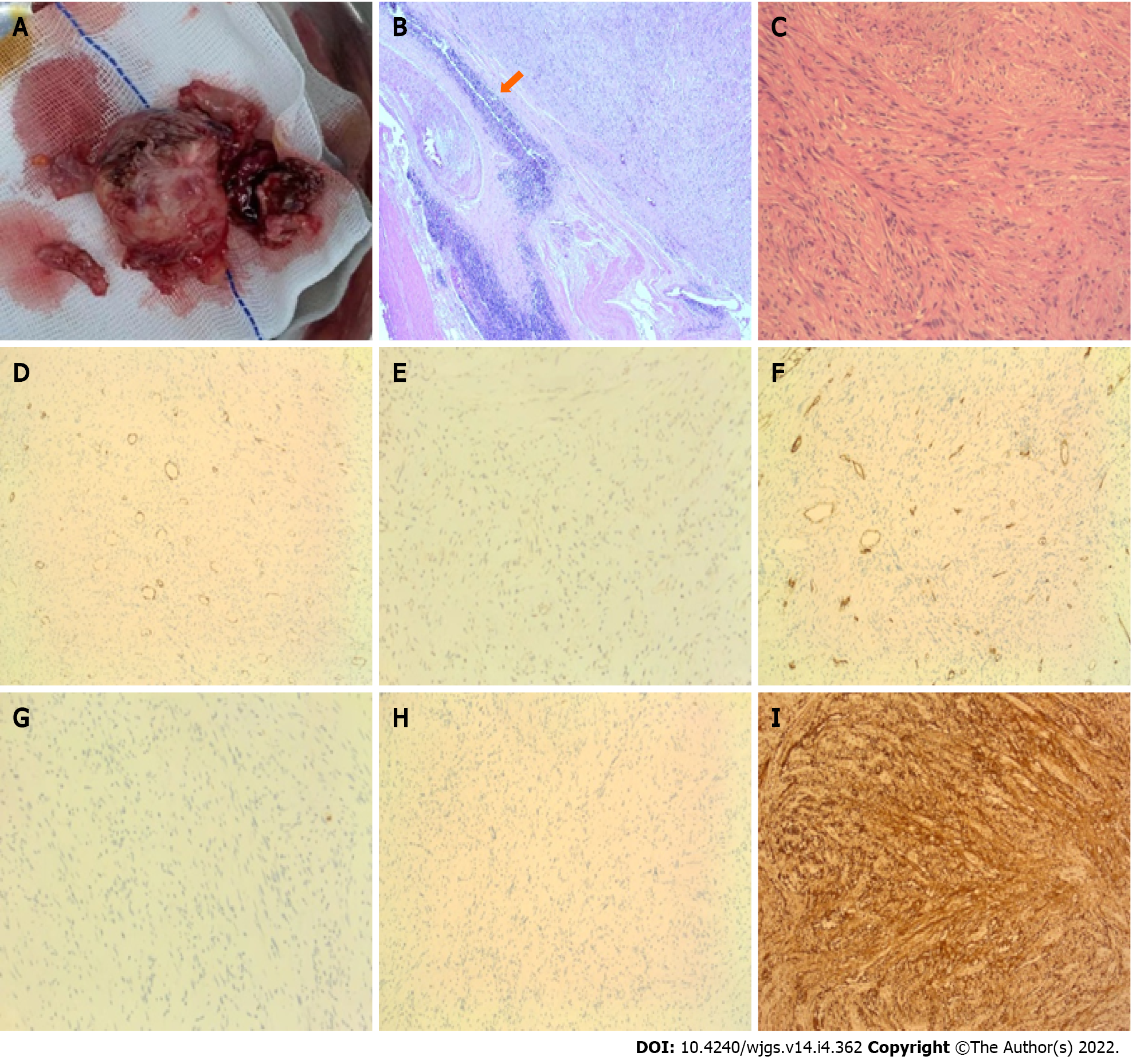
Figure 3 Specimen after surgery, hematoxylin and eosin-stained pathological sections, and immunohistochemistry.
A: The resected tumor; B and C: The tumor comprises intertwined bundles of spindle cells with tapered nuclei; mitotic figures are rare. Lymphocyte infiltration is observed in the tumor tissue, and a characteristic peripheral lymphoid cuff is present (B: 4 × C: 20 ×); D-I: Immunohistochemical staining of the gastric mass confirming a gastric schwannoma with positive staining for S-100 protein (I) and negative staining for α-smooth muscle actin (D), DOG-1 (E), CD34 (F), CD117 (G), and desmin (H).
Figure 4 Timeline of case occurrence.
CT: Computed tomography; EUS: Endoscopic ultrasonography.
- Citation: He CH, Lin SH, Chen Z, Li WM, Weng CY, Guo Y, Li GD. Laparoscopic-assisted endoscopic full-thickness resection of a large gastric schwannoma: A case report. World J Gastrointest Surg 2022; 14(4): 362-369
- URL: https://www.wjgnet.com/1948-9366/full/v14/i4/362.htm
- DOI: https://dx.doi.org/10.4240/wjgs.v14.i4.362


