Copyright
©The Author(s) 2022.
World J Gastrointest Surg. Mar 27, 2022; 14(3): 221-235
Published online Mar 27, 2022. doi: 10.4240/wjgs.v14.i3.221
Published online Mar 27, 2022. doi: 10.4240/wjgs.v14.i3.221
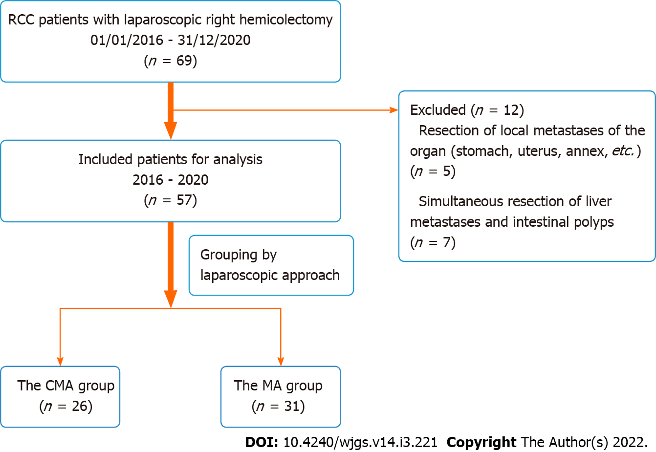
Figure 1 Flow chart of clinical data selection.
Figure 2 The position of the five trocars.
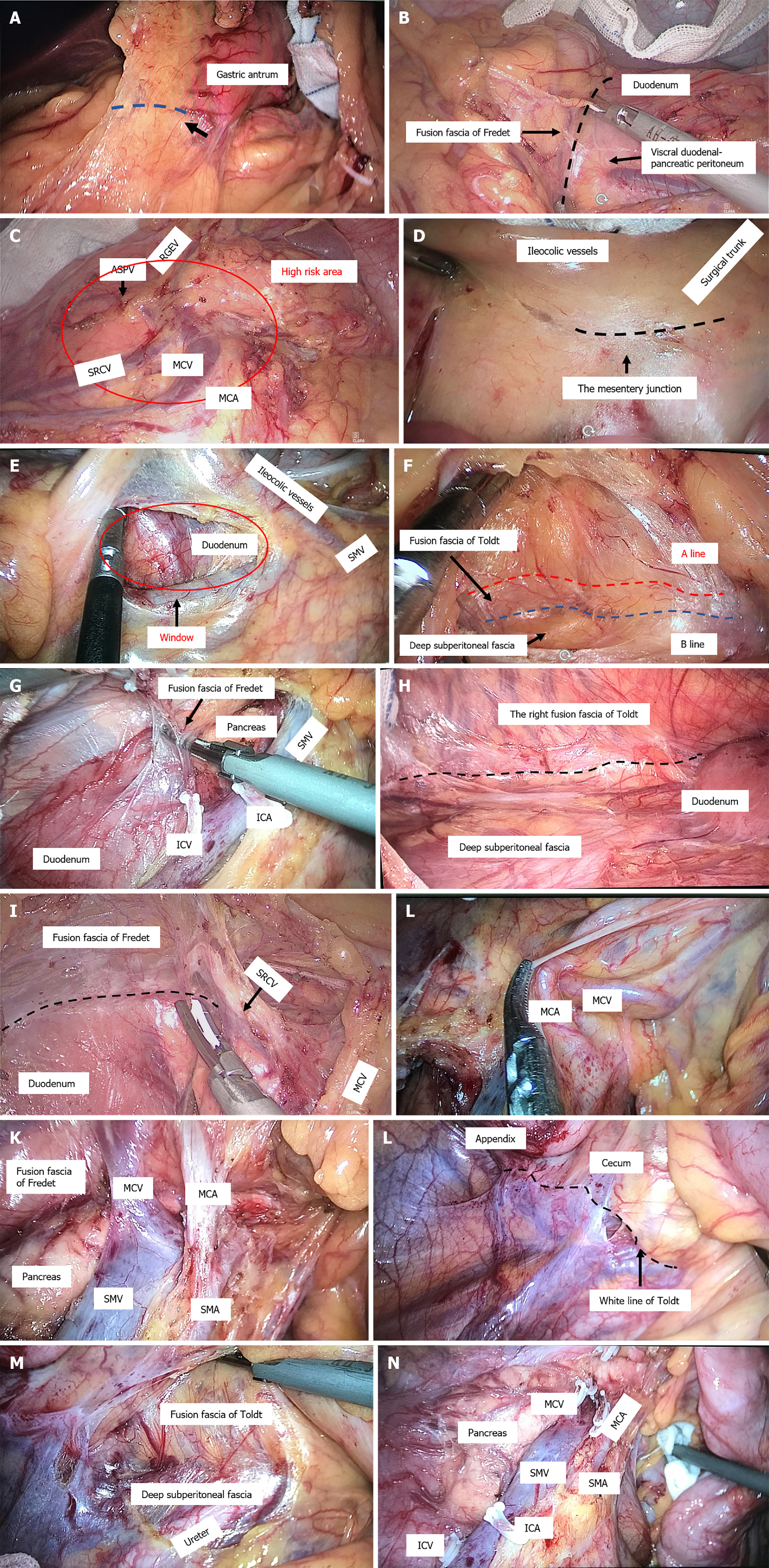
Figure 3 The cranial-medial mixed dominant approach.
A: The right fusion fascia area of the transverse mesocolon and the mesogastrium. The black arrow indicates the position of the first cut with dissection along the dotted line; B: Expanded surgical plane between the fusion fascia of Fredet and the visceral duodenal-pancreatic peritoneum; C: High-risk area using the superior right colic vein as a landmark included the gastrocolic trunk of Henle, middle colic vein (MCV), and middle colic artery (MCA); D: The mesentery junction fusion point of the mesocolon and the intestinal mesentery, approximately 3 cm below the projection of ileocolic vessels to the confluence of the superior mesenteric vein (SMV); E: The mesocolic window was opened to enter the right retrocolic space; F: Expanded surgical plane of the right retrocolic space between the ventral side of the fusion fascia of Toldt and deep subperitoneal fascia. A line: Red dotted line, B line: Blue dotted line, as indicated by Shinohara[15]; G: Fusion fascia of Fredet; H: Right retrocolic space after resection between the fusion fascia of Toldt and deep subperitoneal fascia; I: Rendezvous view of the surgical plane after the cephalic-approach procedure and medial-approach procedure, cut along the black dotted line on the fusion fascia of Fredet; J: Complex three-dimensional anatomical structure of the root of medial colic vessels; K: Three-dimensional dissection of the mesocolon around the root of the MCVs; L: Lateral white line of Toldt around the ileocaecum; M: Cleavage of the lateral white line of Toldt around the caecum connected to the posterior plane of the expanded fusion fascia of Toldt; N: SMV after lymph node dissection. RGEV: Right gastroepiploic vein; ASPV: Anterosuperior pancreatic-duodenal vein; SRCV: Superior right colic vein; ICA: Ileocolic artery; ICV: Ileocolic vein; SMA: Superior mesenteric artery.
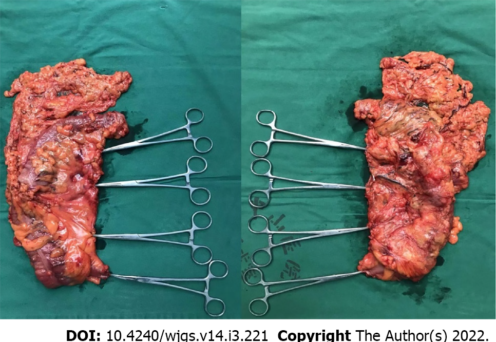
Figure 4 The specimen from the operation.
- Citation: Lin L, Yuan SB, Guo H. Does cranial-medial mixed dominant approach have a unique advantage for laparoscopic right hemicolectomy with complete mesocolic excision? World J Gastrointest Surg 2022; 14(3): 221-235
- URL: https://www.wjgnet.com/1948-9366/full/v14/i3/221.htm
- DOI: https://dx.doi.org/10.4240/wjgs.v14.i3.221


