Copyright
©The Author(s) 2022.
World J Gastrointest Surg. Dec 27, 2022; 14(12): 1432-1437
Published online Dec 27, 2022. doi: 10.4240/wjgs.v14.i12.1432
Published online Dec 27, 2022. doi: 10.4240/wjgs.v14.i12.1432
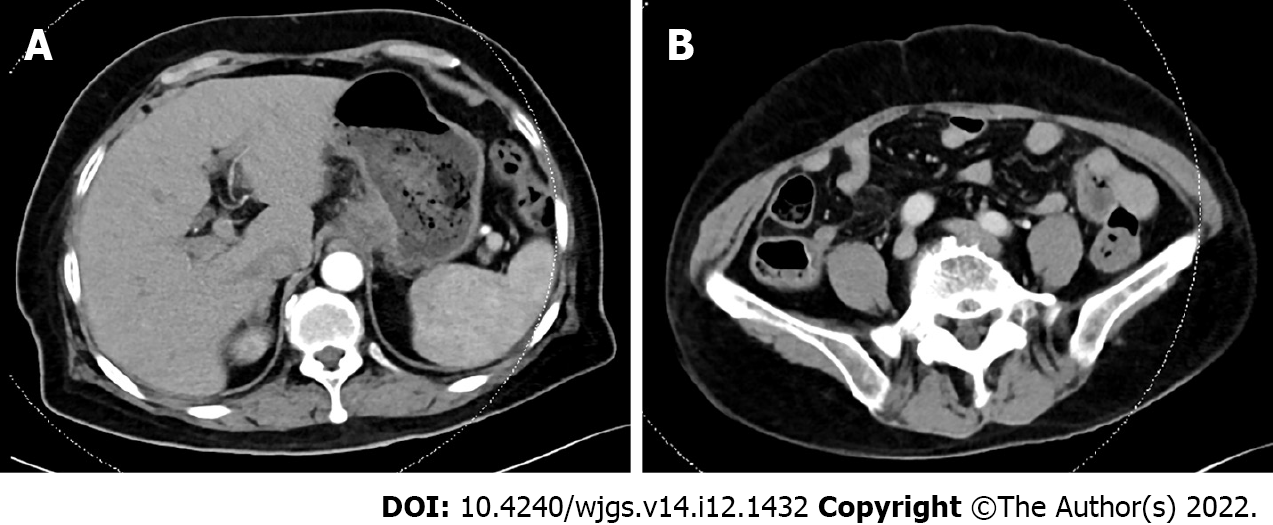
Figure 1 Imaging data of May 23, 2022.
A: Computed tomography (CT) showed that the cardia was thickened and reinforced; B: CT showed that the appendix was normal.
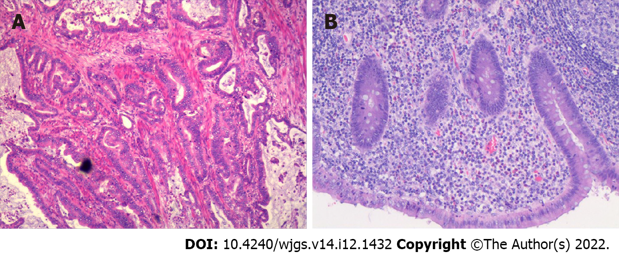
Figure 2 Photomicrographs (hematoxylin and eosin, × 200 magnification).
A: Moderately differentiated tubular adenocarcinoma; B: Acute purulent appendicitis and periapical inflammation.
Figure 3 Imaging data of June 4, 2022.
A: Computed tomography showed no abnormalities around the anastomosis or stump, no leakage into the abdominal cavity from oral pantothenic glucosamine, no abnormalities in the ileocecal region or appendix, and no significant fluid accumulation in the abdominopelvic cavity; B: No abnormalities were found in the ileocecal region or duodenal stump; C: No abnormalities were found in the appendix.
Figure 4 Imaging data of June 10, 2022.
A: Computed tomography showed that there was effusion in the perihepatic and hepatorenal interstitial areas; B: There was effusion in the right paracolic sulcus and exudative changes in the right iliac fossa; C: Effusion was seen in the pelvis.
Figure 5 laparoscopic exploration.
A and B: Yellow-green fluid could be found in the perihepatic and pelvic cavities; C: The duodenal stump was wrapped in tissue and did not show leakage.
Figure 6 Surgical procedure.
A: A large amount of pus moss was seen around the ileocecal region, and the terminal ileum wrapped around the appendix; B: A septic and swollen appendix was seen after careful laparoscopic separation of the adhesions (white arrow); C: Resected appendix (red arrow) and treated appendiceal stump and mesentery.
Figure 7 Imaging data of July 12, 2022.
Computed tomography showed no significant abnormalities of the duodenal stump, appendiceal stump or pelvic. A: Duodenal stump; B: Appendiceal stump; C: Pelvic.
- Citation: Ma J, Zha ZP, Zhou CP, Miao X, Duan SQ, Zhang YM. Acute appendicitis in the short term following radical total gastrectomy misdiagnosed as duodenal stump leakage: A case report. World J Gastrointest Surg 2022; 14(12): 1432-1437
- URL: https://www.wjgnet.com/1948-9366/full/v14/i12/1432.htm
- DOI: https://dx.doi.org/10.4240/wjgs.v14.i12.1432


