Copyright
©The Author(s) 2021.
World J Gastrointest Surg. Sep 27, 2021; 13(9): 988-999
Published online Sep 27, 2021. doi: 10.4240/wjgs.v13.i9.988
Published online Sep 27, 2021. doi: 10.4240/wjgs.v13.i9.988
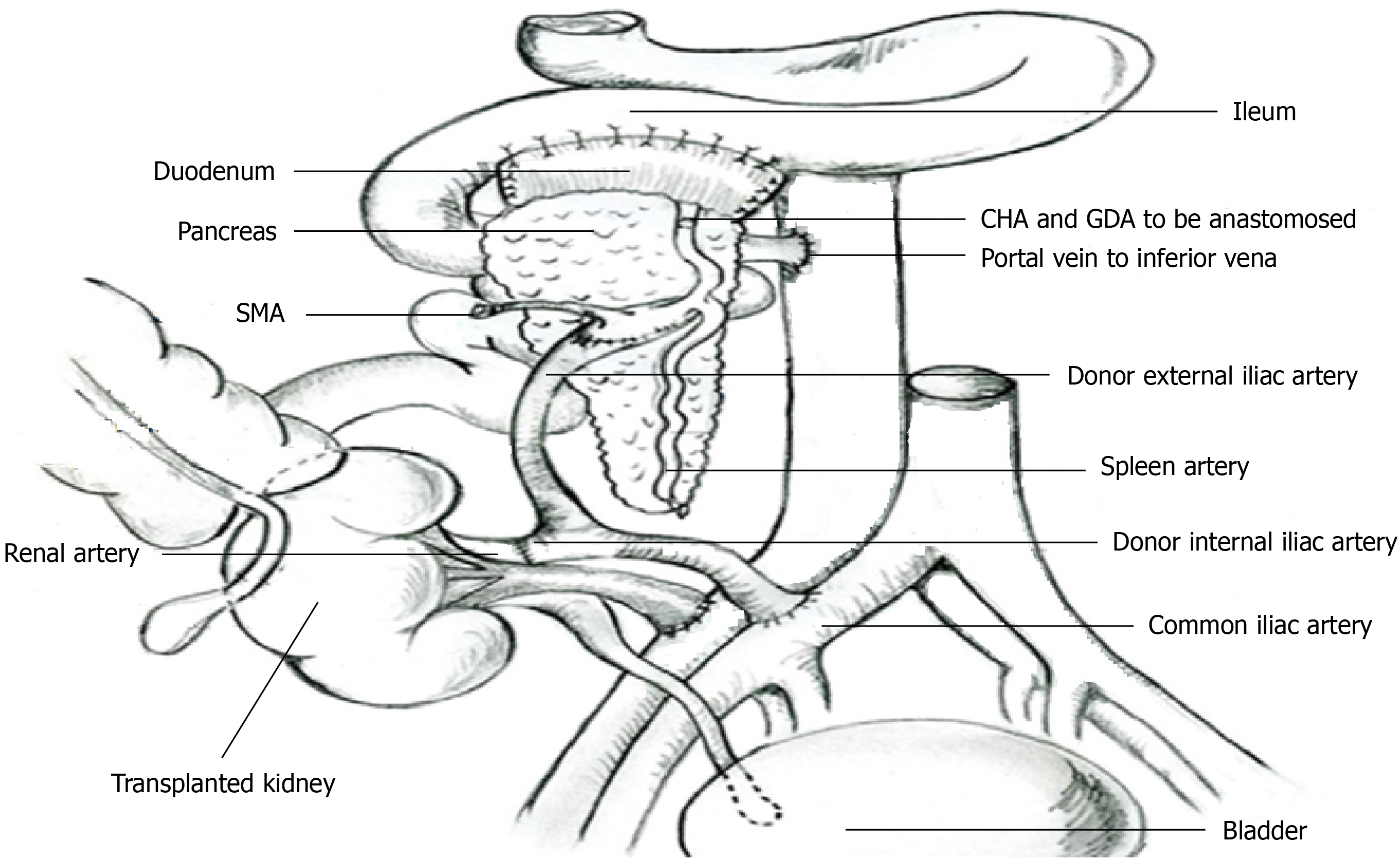
Figure 1 Revascularization of the pancreas and kidney with a single arterial conduit.
The donor duodenum segment was anastomosed side-to-side to the recipient’s distal ileum. CHA: Common hepatic artery; GDA: Gastroduodenal arterial; SMA: Superior mesenteric artery.
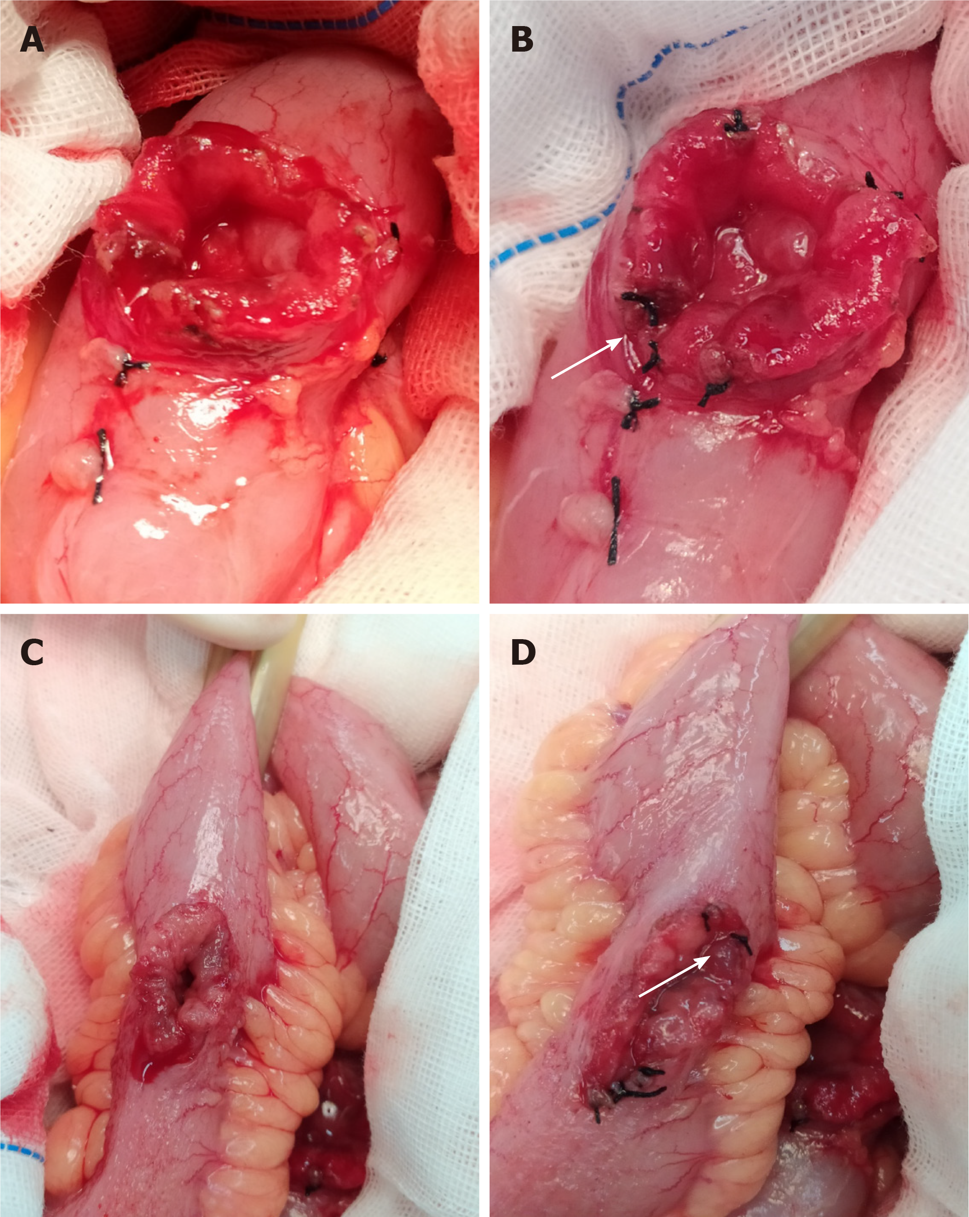
Figure 2 Suture ligation for submucosal hemostasis.
A: Bleeding at the cut edge of the duodenum; B: Bleeding spots of the duodenum were staunched by transmural suture ligation; C: Bleeding at the cut edge of the ileum; D: Bleeding spots of the ileum were staunched by transmural suture ligation. White arrow: Knot of suture thread.
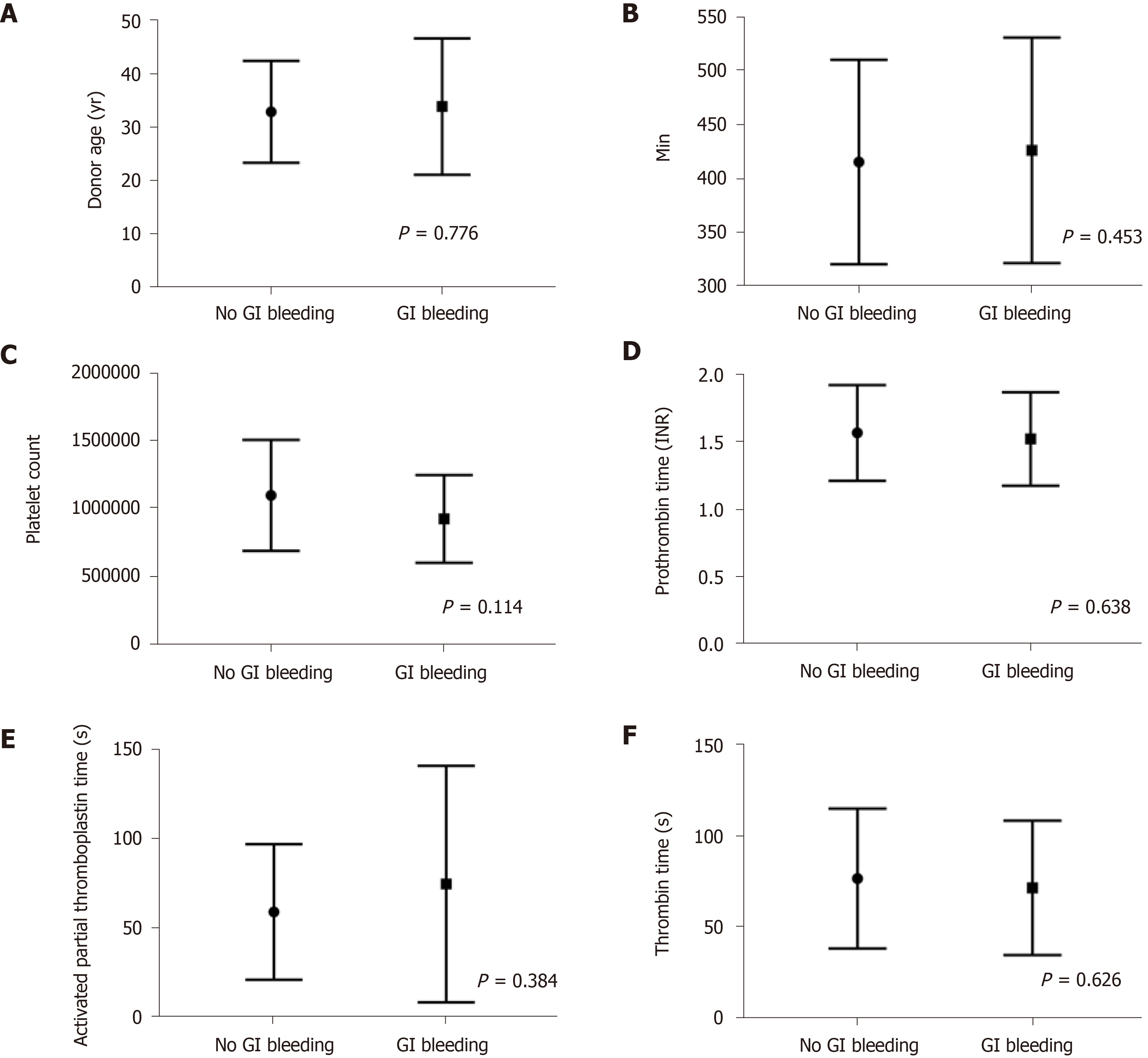
Figure 3 Comparison of clinical features within 1 wk postoperatively in gastrointestinal bleeding and no gastrointestinal bleeding group.
A: Donor age; B: Mean pancreatic graft cold ischemia time; C: Platelet count; D: Prothrombin time international normalized rate; E: Activated partial thromboplastin time; F: Thrombin time. GI: Gastrointestinal; INR: International normalized rate.
Figure 4 The Kaplan–Meier curves for patient, kidney graft, and pancreas graft in suture ligation and no suture ligation group.
A: Patient survival curves; B: Kidney graft survival curves; C: Pancreas graft survival curves. SL: Suture ligation; NSL: No suture ligation.
- Citation: Wang H, Fu YX, Song WL, Mo CB, Feng G, Zhao J, Pei GH, Shi XF, Wang Z, Cao Y, Nian YQ, Shen ZY. Suture ligation for submucosal hemostasis during hand-sewn side-to-side duodeno-ileostomy in simultaneous pancreas and kidney transplantation. World J Gastrointest Surg 2021; 13(9): 988-999
- URL: https://www.wjgnet.com/1948-9366/full/v13/i9/988.htm
- DOI: https://dx.doi.org/10.4240/wjgs.v13.i9.988


