Copyright
©The Author(s) 2021.
World J Gastrointest Surg. Sep 27, 2021; 13(9): 1102-1109
Published online Sep 27, 2021. doi: 10.4240/wjgs.v13.i9.1102
Published online Sep 27, 2021. doi: 10.4240/wjgs.v13.i9.1102
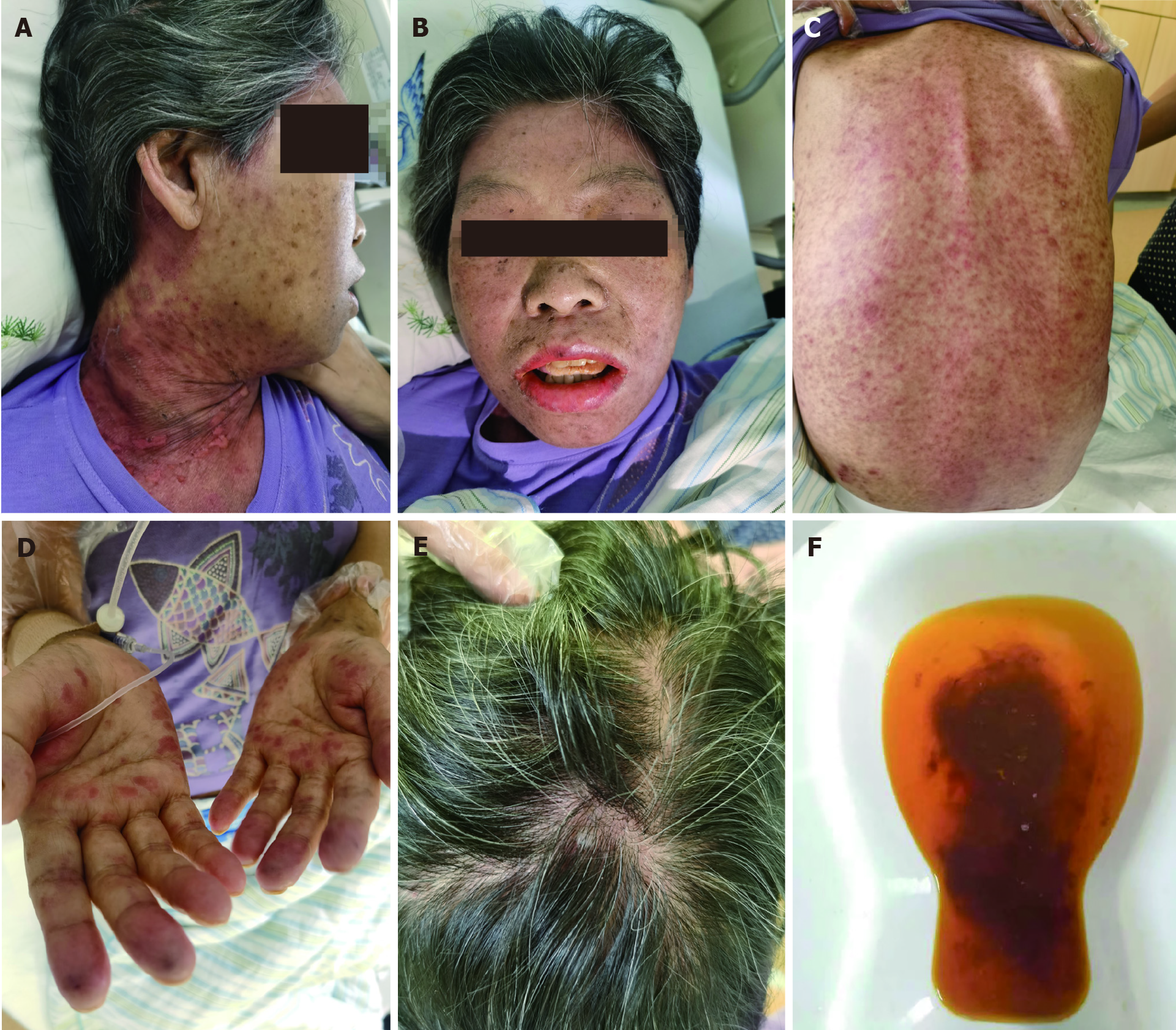
Figure 1 Clinical ground observations of graft-vs-host disease.
A: Anterior cervical rash on post-operative day 24; B: Oral ulcers on post-operative day 25; C: Dorsal rash on post-operative day 25; D and E: Palmar rash on post-operative day 22; F: Scalp rash on post-operative day 27; G: Passage of three or more loose or liquid stools per day.
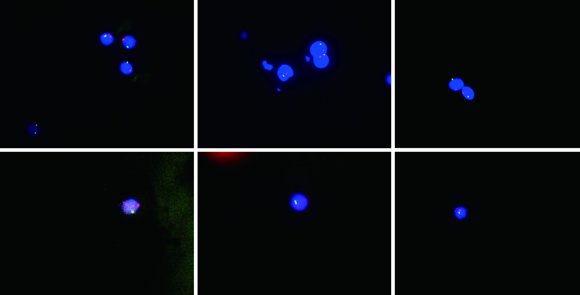
Figure 2 Twelve erythrocytes analyzed, 11 showed an XY signal pattern, while one showed an XX signal pattern (91.
7% showed one X and one Y signal, and 8.3% showed two X signals). Y is the red fluorescent signal; X is the green fluorescent signal.
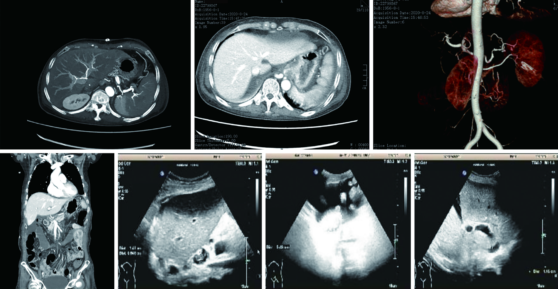
Figure 3
The portal vein, inferior vena cava, hepatic artery, and hepatic venous blood flow were smooth.
- Citation: Xiao JJ, Ma JY, Liao J, Wu D, Lv C, Li HY, Zuo S, Zhu HT, Gu HJ. Fluorescence in situ hybridization-based confirmation of acute graft-vs-host disease diagnosis following liver transplantation: A case report. World J Gastrointest Surg 2021; 13(9): 1102-1109
- URL: https://www.wjgnet.com/1948-9366/full/v13/i9/1102.htm
- DOI: https://dx.doi.org/10.4240/wjgs.v13.i9.1102


