Copyright
©The Author(s) 2021.
World J Gastrointest Surg. Sep 27, 2021; 13(9): 1095-1101
Published online Sep 27, 2021. doi: 10.4240/wjgs.v13.i9.1095
Published online Sep 27, 2021. doi: 10.4240/wjgs.v13.i9.1095
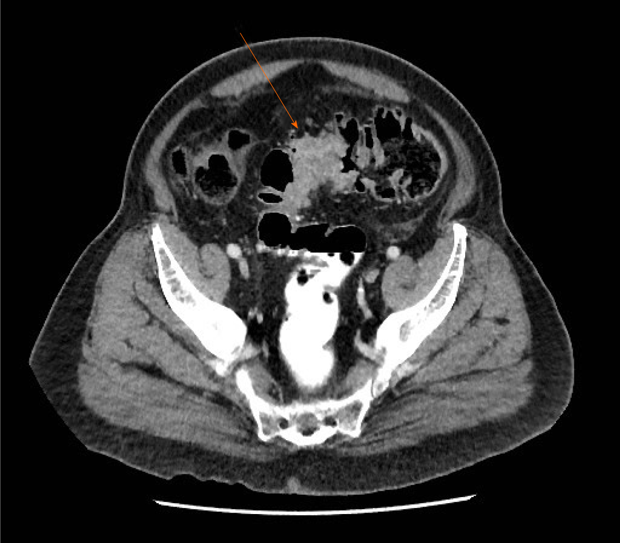
Figure 1 Axial contrast-enhanced computed tomography image.
A short segment circumferential soft tissue mass within the sigmoid colon and luminal narrowing (arrow) consistent with a tumor. There is a small lymph node adjacent to the lesion.
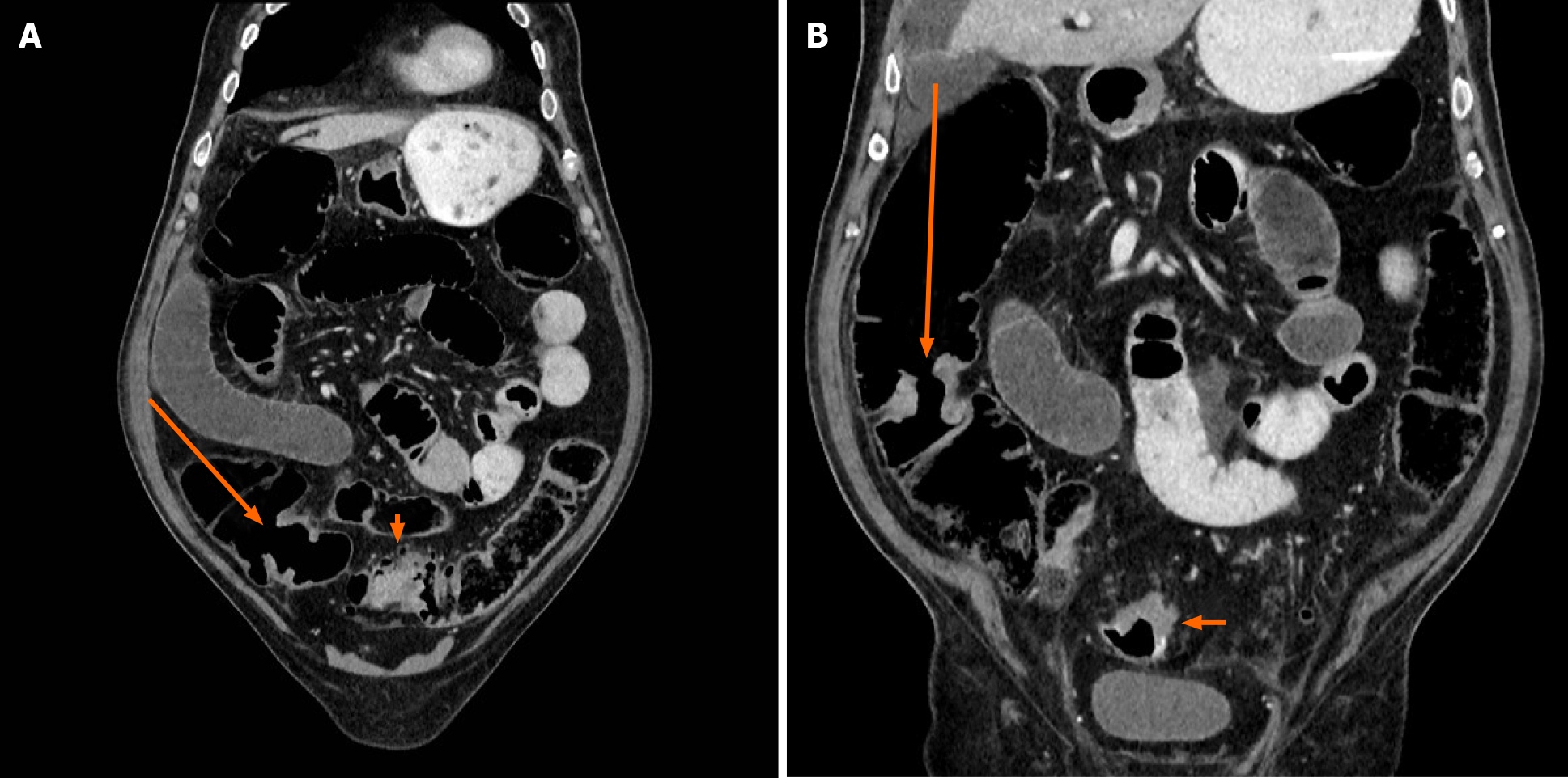
Figure 2 Coronal contrast-enhanced computed tomography image.
A: The proximal right colonic tumor (long arrow) at the level of the ileocecal valve, evidenced by a focal mild circumferential wall thickening. Sigmoid cancer is partially seen (short arrow); B: The middle right colonic tumor (long arrow), evidenced by a focal circumferential wall thickening without obstruction. Sigmoid cancer is partially seen (short arrow).
- Citation: Bergeron E, Maniere T, Do XV, Bensoussan M, De Broux E. Three colonic cancers, two sites of complete occlusion, one patient: A case report. World J Gastrointest Surg 2021; 13(9): 1095-1101
- URL: https://www.wjgnet.com/1948-9366/full/v13/i9/1095.htm
- DOI: https://dx.doi.org/10.4240/wjgs.v13.i9.1095


