Copyright
©The Author(s) 2021.
World J Gastrointest Surg. Aug 27, 2021; 13(8): 772-787
Published online Aug 27, 2021. doi: 10.4240/wjgs.v13.i8.772
Published online Aug 27, 2021. doi: 10.4240/wjgs.v13.i8.772
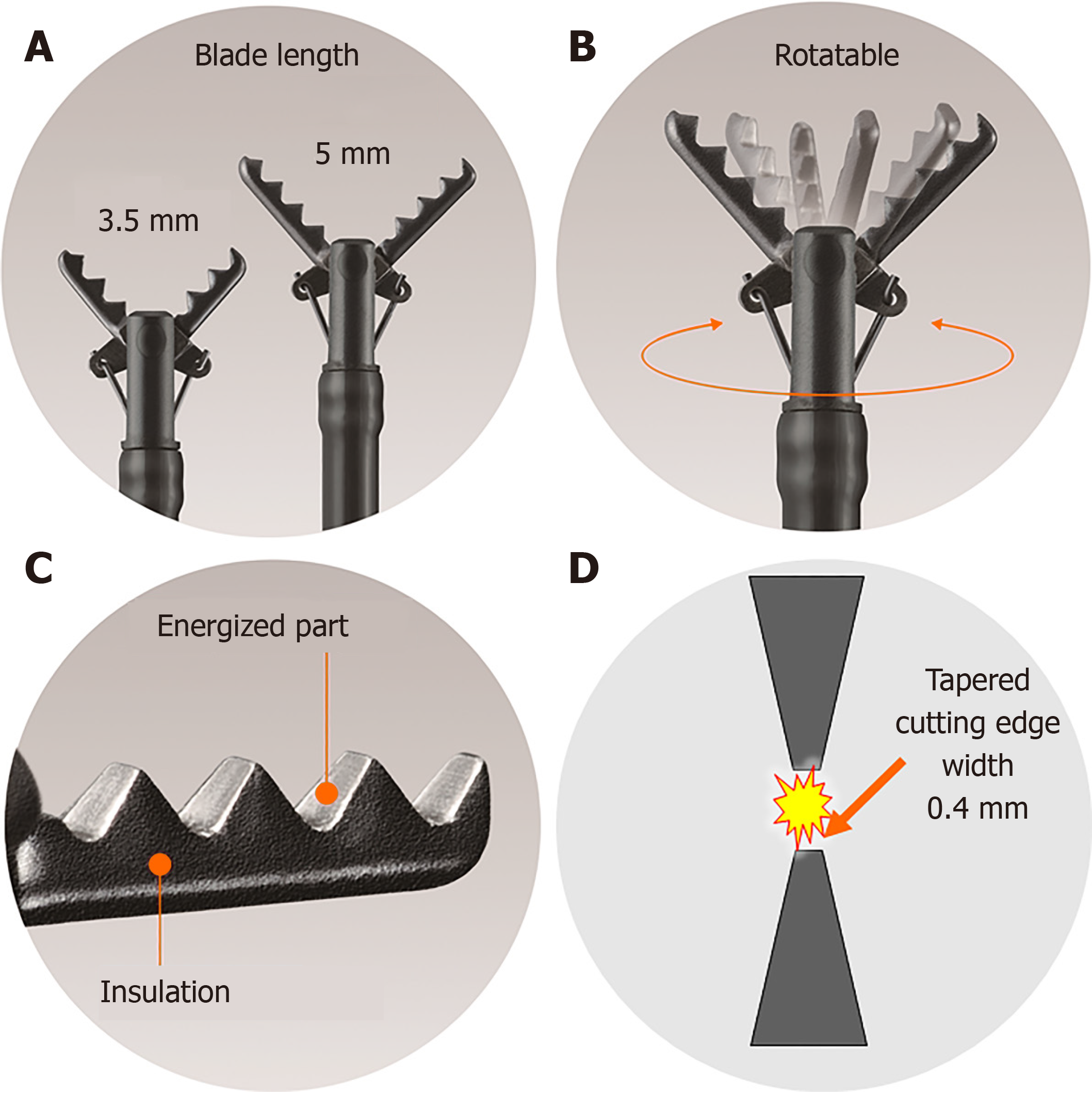
Figure 1 Mechanical features of the clutch cutter.
A: Two blade lengths (5 mm and 3.5 mm) are available. The blade is saw-toothed over the entire length of the scissors; B: Rotatable. The blade part can rotate 360°; C: The cutting edge is serrated, and all surfaces other than the cutting edge are insulated; D: Sectional view of the cutting edge. The cutting edge is tapered to a width of 0.4 mm, which is the same width as the polypectomy snare, so that the tissue can be pressed and cut. Quoted and modified from Akahoshi et al[16] and Fujifilm (https://asset.fujifilm.com/master/jp/files/2020-07/bbd3e2ffc64eb7d6cd4e135f80339d46/endoscopy-esd-clutchcutter-catalog.pdf) with permission. Citation: Akahoshi K, Kubokawa M, Tamura S. Clutch Cutter (Long type & Short type). In: Yahagi N. Electrosurgical units for therapeutic endoscopy-Settings and Its effective usage. 3rd ed. Tokyo: Japan Medical Center, 2020: 177-184.
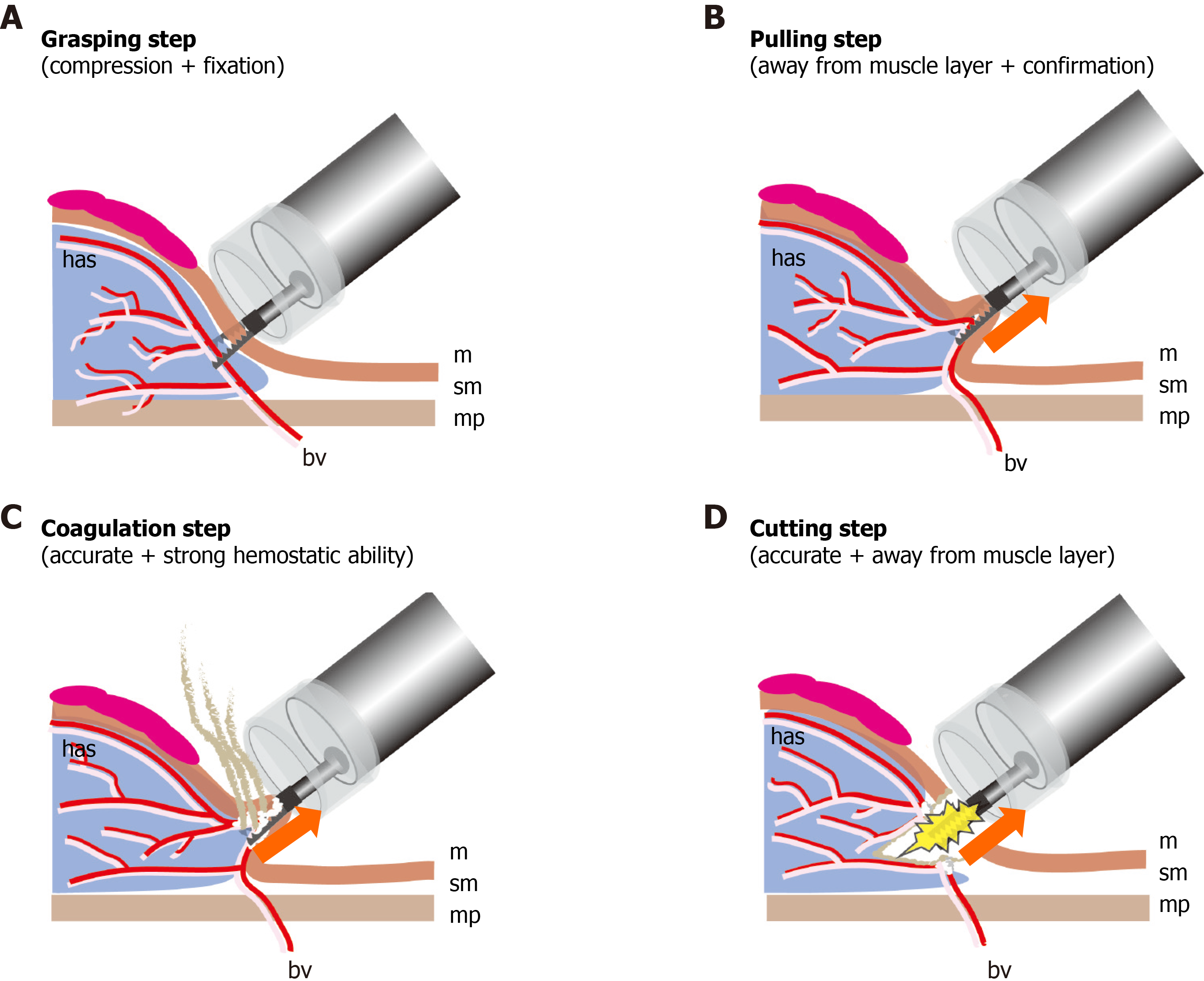
Figure 2 Four basic steps for safe and effective endoscopic submucosal dissection using the clutch cutter.
A: Grasping step (lock-on-like accurate targeting); B: Pulling step (minimal electrical damage to the muscle layer); C: Coagulation step (accurate and effective hemostatic ability); D: Cutting step (accurate mucosal incision and submucosal dissection). Quoted and modified from Akahoshi et al[16] with permission. Citation: Akahoshi K, Kubokawa M, Tamura S. Clutch Cutter (Long type & Short type). In: Yahagi N. Electrosurgical units for therapeutic endoscopy-Settings and Its effective usage. 3rd ed. Tokyo: Japan Medical Center, 2020: 177-184.
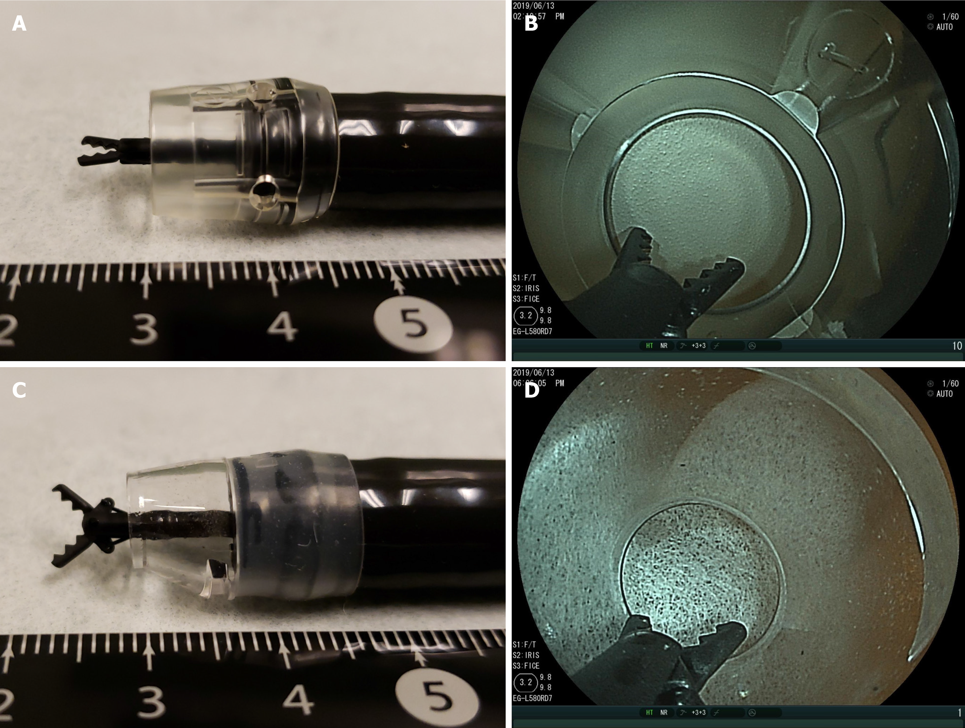
Figure 3 Hoods for endoscopic submucosal dissection using the clutch cutter.
A: Overview of the long transparent hood (F-01; TOP Corp., Tokyo, Japan); B: Internal field of view with the long transparent hood (clutch cutter long type, half-open state); C: Overview of the tapered transparent hood (ST hood long type, DH-40GR; Fujifilm, Tokyo); D: Internal field of view of the tapered transparent hood (clutch cutter short-type, half-open state). Quoted and modified from Akahoshi et al[16] with permission. Citation: Akahoshi K, Kubokawa M, Tamura S. Clutch Cutter (Long type & Short type). In: Yahagi N. Electrosurgical units for therapeutic endoscopy-Settings and Its effective usage. 3rd ed. Tokyo: Japan Medical Center, 2020: 177-184.
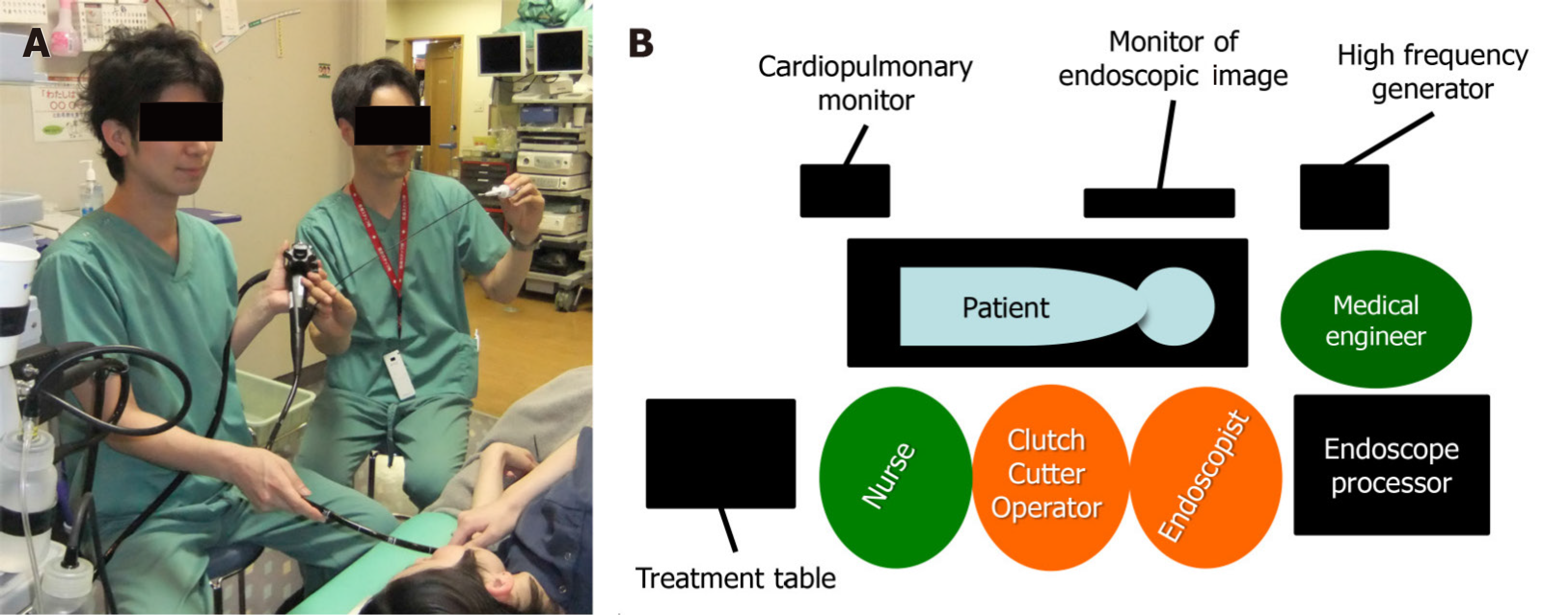
Figure 4 Equipment and personnel allocation in endoscopic submucosal dissection using the clutch cutter.
Quoted and modified from Akahoshi et al[17] with permission. Citation: Akahoshi K, Motomura Y, Kubokawa M, Kinoshita N. Characteristics and effective use of the Clutch Cutter. Endoscopia Digestiva 2014; 26: 1399-1406.
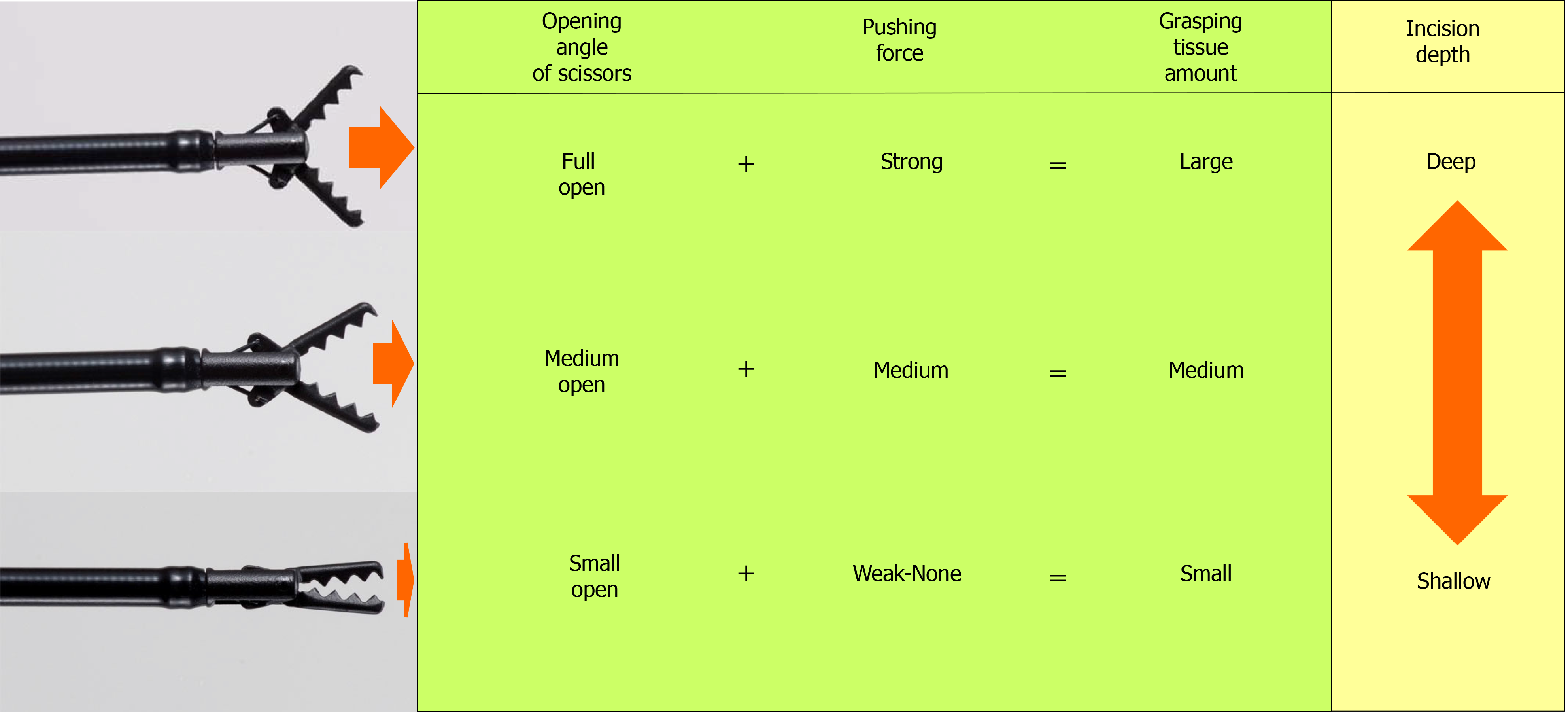
Figure 5 Adjusting the incision depth with the clutch cutter.
Quoted and modified from Akahoshi et al[16] with permission. Citation: Akahoshi K, Kubokawa M, Tamura S. Clutch Cutter (Long type & Short type). In: Yahagi N. Electrosurgical units for therapeutic endoscopy-Settings and Its effective usage. 3rd ed. Tokyo: Japan Medical Center, 2020: 177-184.
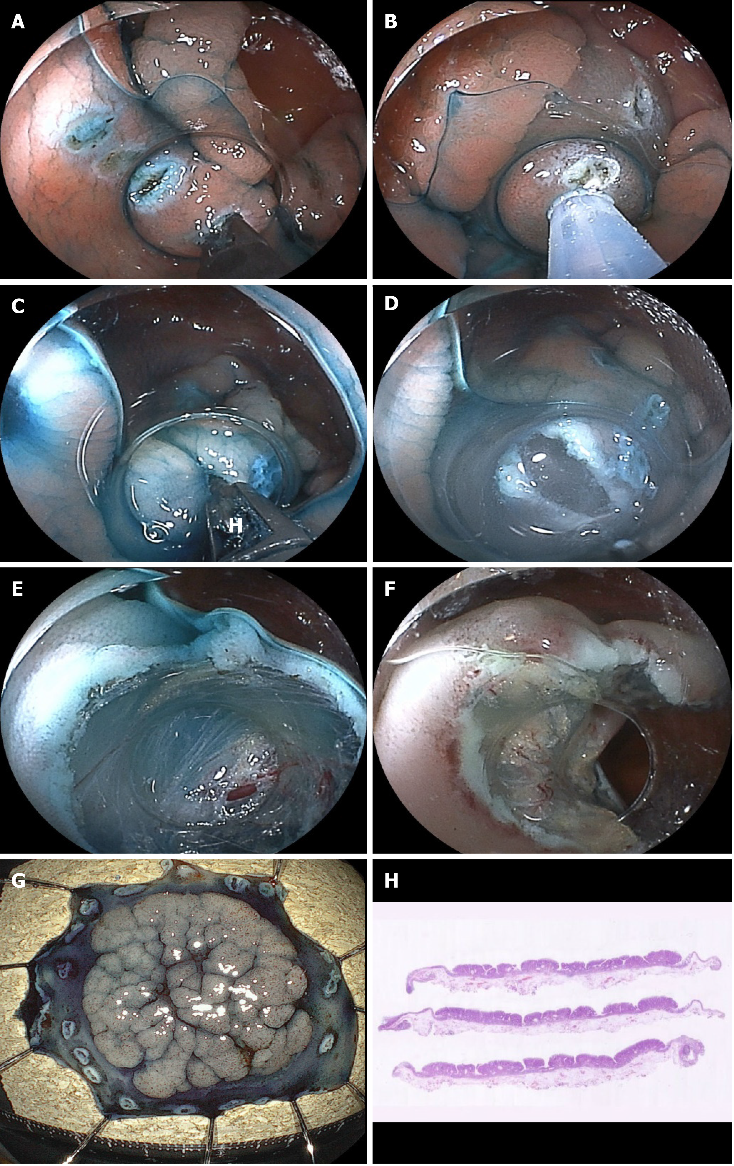
Figure 6 Endoscopic view of each step of endoscopic submucosal dissection using the clutch cutter for granular and homogeneous type laterally spreading tumor of the ascending colon.
A: Several points along the contour of the lesion are marked with a coagulation current; B: Submucosal injection of hyaluronic acid solution; C: The targeted mucosa is grasped outside the marking points for fixation prior to first mucosal incision; D: First mucosal incision; E: The deep submucosal layer is gradually dissected using the clutch cutter from the muscularis propria; F: The tumor is completely resected; G: The resected specimen shows en-bloc resection of the tumor; H: Cross-section of the resected specimen showing R0 resection.
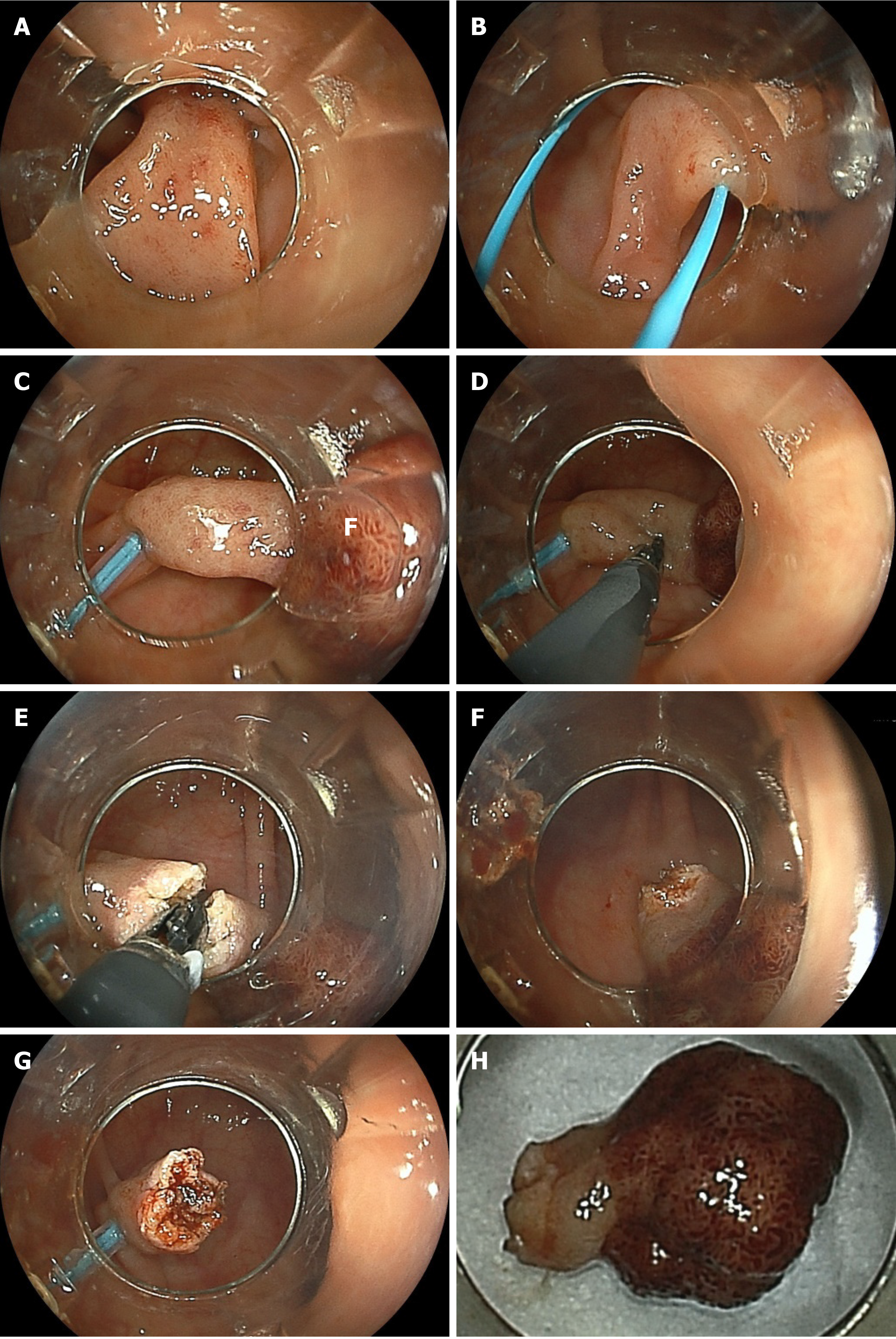
Figure 7 Endoscopic polypectomy using the clutch cutter and the detachable snare for large pedunculated colonic polyp.
A: Long and thick stalk of the sigmoid colonic polyp; B: Positioning the detachable snare at the base of the stalk; C: Squeezing the thick stalk using a detachable snare; D: The Clutch Cutter is grasping the stalk 10mm above the detachable snare under direct endoscopic view; E: The Clutch Cutter is cutting the stalk using electrosurgical current; F: The polyp is removed without bleeding; G: Enough distance between the excised margin and the detachable snare; H: The resected specimen of the polyp.
- Citation: Akahoshi K, Komori K, Akahoshi K, Tamura S, Osada S, Shiratsuchi Y, Kubokawa M. Advances in endoscopic therapy using grasping-type scissors forceps (with video). World J Gastrointest Surg 2021; 13(8): 772-787
- URL: https://www.wjgnet.com/1948-9366/full/v13/i8/772.htm
- DOI: https://dx.doi.org/10.4240/wjgs.v13.i8.772


