Copyright
©The Author(s) 2021.
World J Gastrointest Surg. May 27, 2021; 13(5): 406-418
Published online May 27, 2021. doi: 10.4240/wjgs.v13.i5.406
Published online May 27, 2021. doi: 10.4240/wjgs.v13.i5.406
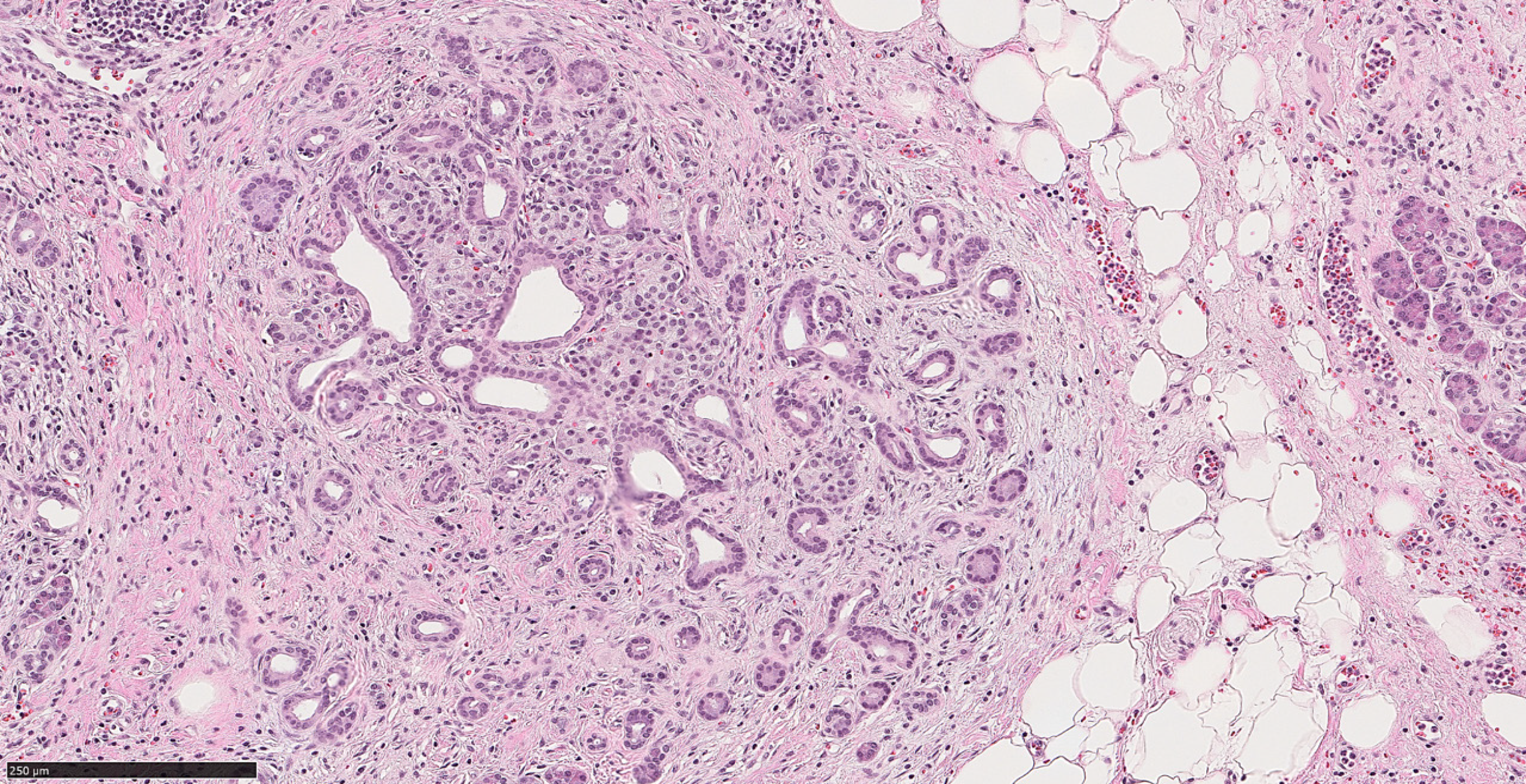
Figure 1 Atrophic glands in a background of chronic pancreatitis (hematoxylin & eosin, 150 ×, scale 250 µm).
In a biopsy material it can be challenging to distinguish this morphology from well differentiated pancreatic ductal adenocarcinoma.
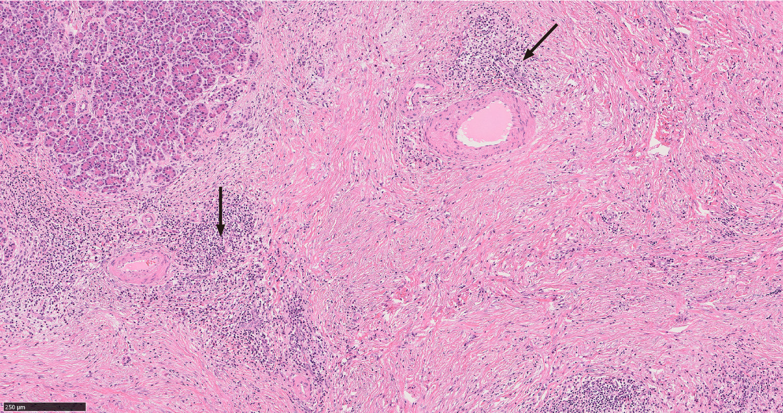
Figure 2 Autoimmune pancreatitis type 1.
This image shows characteristic histologic features of this entity: lymphoplasmacytic infiltration, storiform fibrosis and obliterative phlebitis (arrows) (hematoxylin & eosin, 100 ×, scale 250 µm).
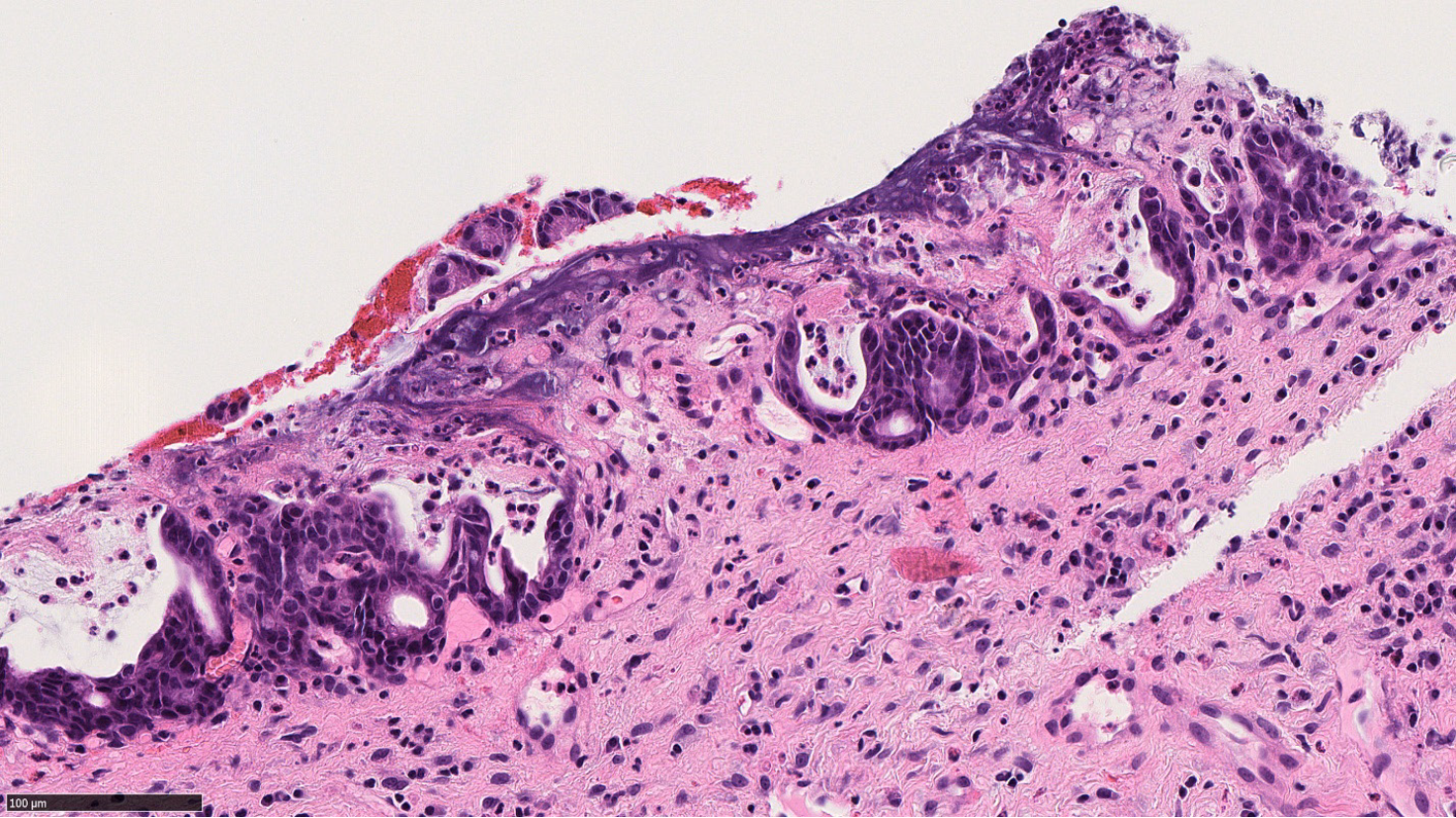
Figure 3 Autoimmune pancreatitis type 2.
This image shows granulocytic infiltration of pancreatic ductal epithelium (hematoxylin & eosin, 300 ×, scale 100 µm).
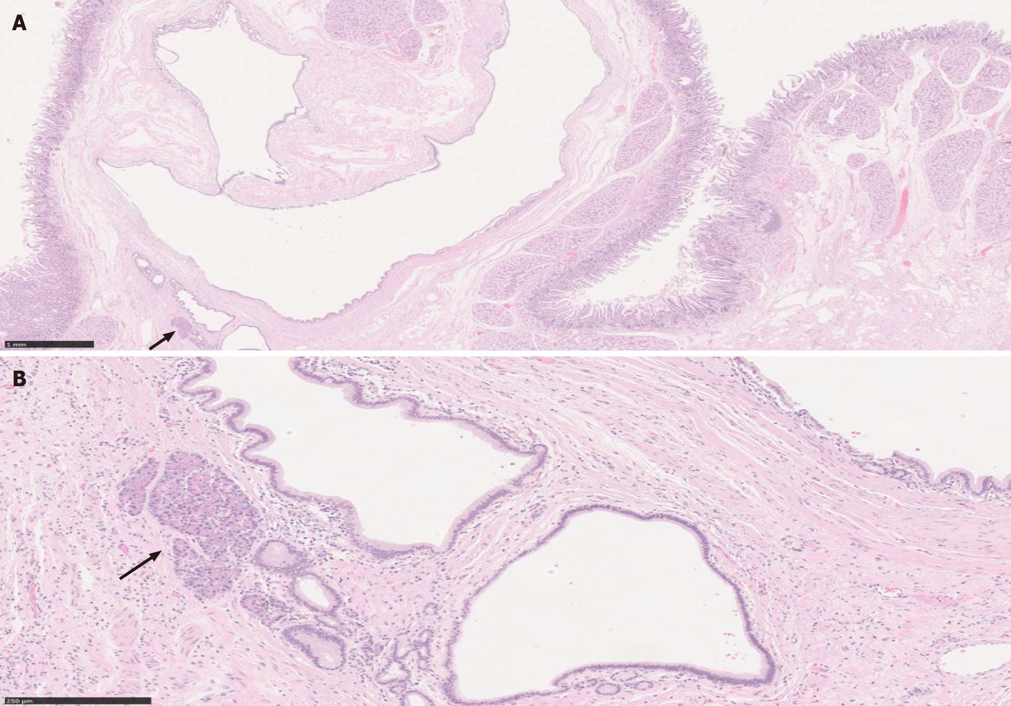
Figure 4 Paraduodenal pancreatitis.
A: This image shows cyst formation, Brunner gland hyperplasia, and ectopic pancreatic tissue (solid arrow) with associated inflammation, consistent with paraduodenal pancreatitis, in a patient with history of significant alcohol use [hematoxylin & eosin (H&E), 20 ×, scale 1 mm]; B: A higher magnification to show the ectopic pancreatic tissue in association with other elements (H&E, 130 ×, scale 250 µm).
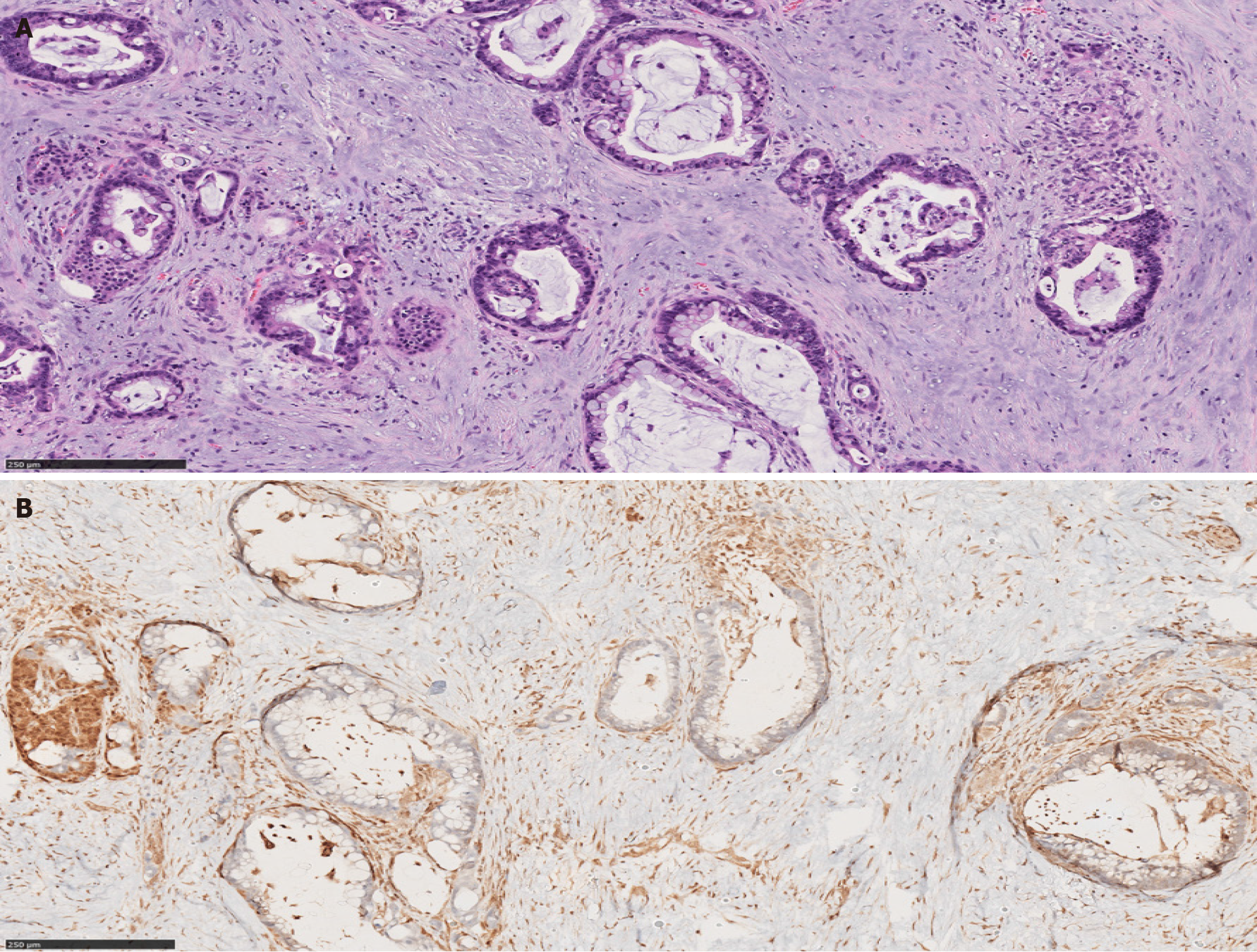
Figure 5 Pancreatic ductal adenocarcinoma.
A: The ducts show features of malignancy including cytologic atypia and incomplete gland formation with loss of lobular architecture, in a prominent desmoplastic stroma (hematoxylin & eosin, 130 ×, scale 250 µm); B: SMAD4 immunostaining shows loss of SMAD4 expression in the malignant glands (SMAD4, 120 ×, scale 250 µm).
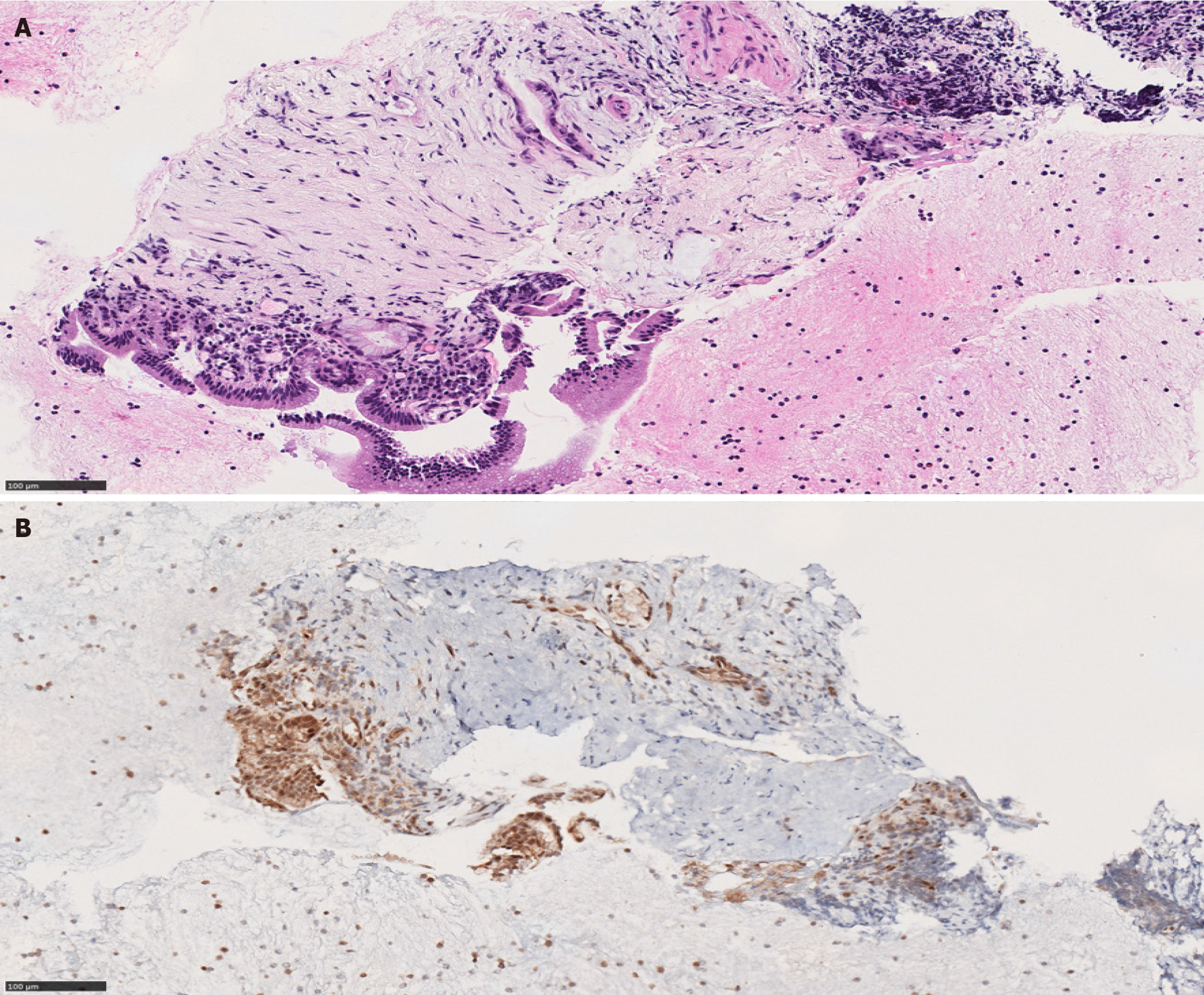
Figure 6 Pancreatic mass biopsy showing a minute focus of atypical glandular structure, highly suspicious for carcinoma.
A: The atypical glands are surrounded by desmoplastic stromal reaction (hematoxylin & eosin, 190 ×, scale 100 µm); B: The atypical glands retain SMAD4 expression; this staining pattern does not rule out malignancy (SMAD4, 190 ×, scale 100 µm).
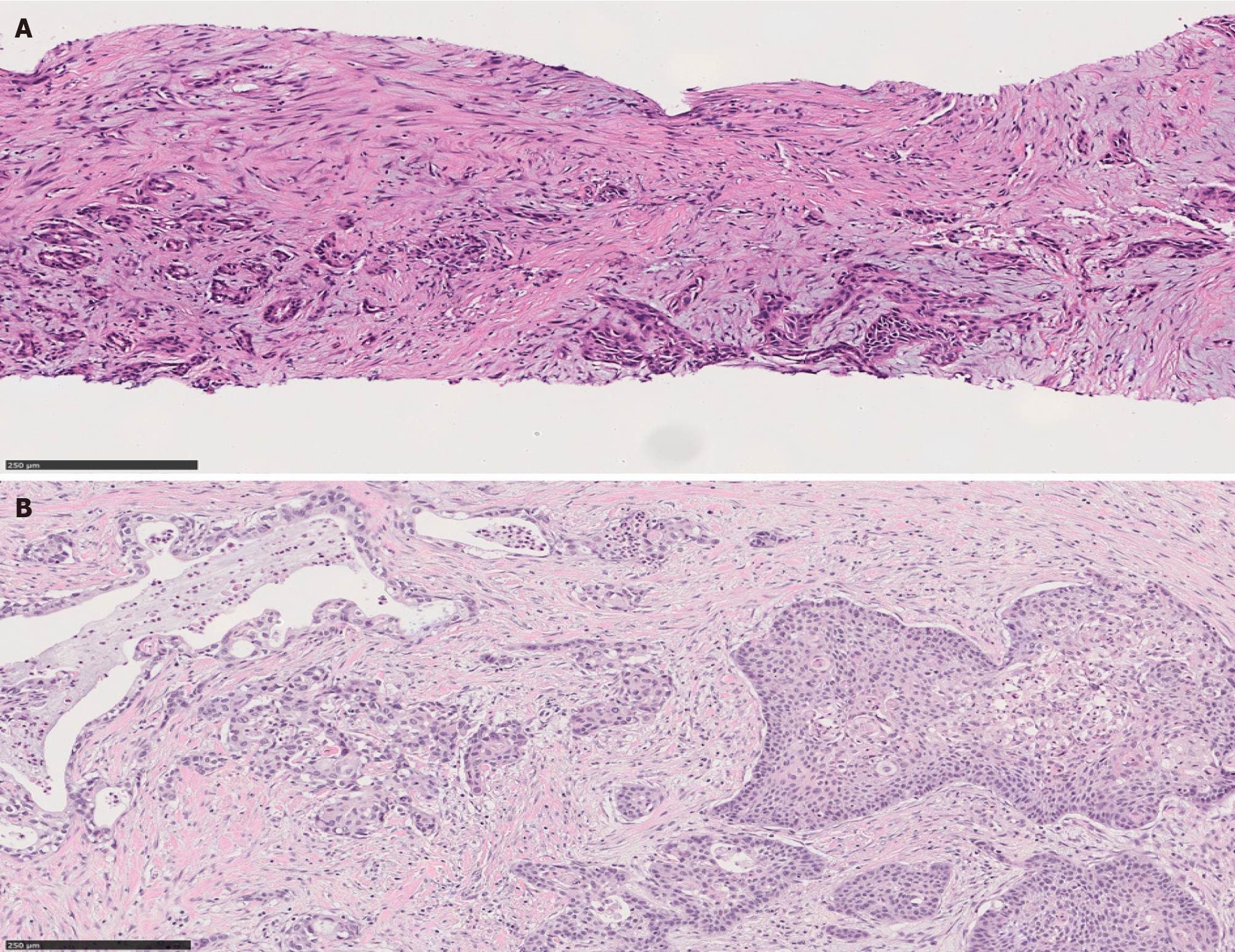
Figure 7 Adenosquamous carcinoma of the pancreas.
A: Adenosquamous carcinoma of the pancreas in a biopsy material. Both squamous component (right) and glandular component (left) are found in this field [hematoxylin & eosin (H&E), 140 ×, scale 250 µm]; B: Adenosquamous carcinoma of the pancreas in a resection material. Squamous nests are noted (right). The glandular counterpart shows typical features of pancreatic ductal adenocarcinoma (left) (H&E, 136 ×, scale 250 µm).
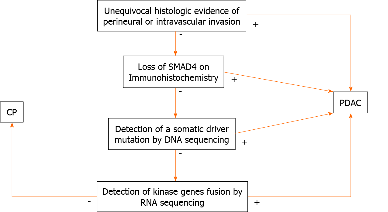
Figure 8 An algorithm outlining the steps for the microscopic, immunohistochemical and molecular work up of pancreatic ductal adenocarcinoma vs chronic pancreatitis.
CP: Chronic pancreatitis; PDAC: Pancreatic ductal adenocarcinoma.
- Citation: Aldyab M, El Jabbour T, Parilla M, Lee H. Benign vs malignant pancreatic lesions: Molecular insights to an ongoing debate. World J Gastrointest Surg 2021; 13(5): 406-418
- URL: https://www.wjgnet.com/1948-9366/full/v13/i5/406.htm
- DOI: https://dx.doi.org/10.4240/wjgs.v13.i5.406


