Copyright
©The Author(s) 2021.
World J Gastrointest Surg. Apr 27, 2021; 13(4): 355-365
Published online Apr 27, 2021. doi: 10.4240/wjgs.v13.i4.355
Published online Apr 27, 2021. doi: 10.4240/wjgs.v13.i4.355
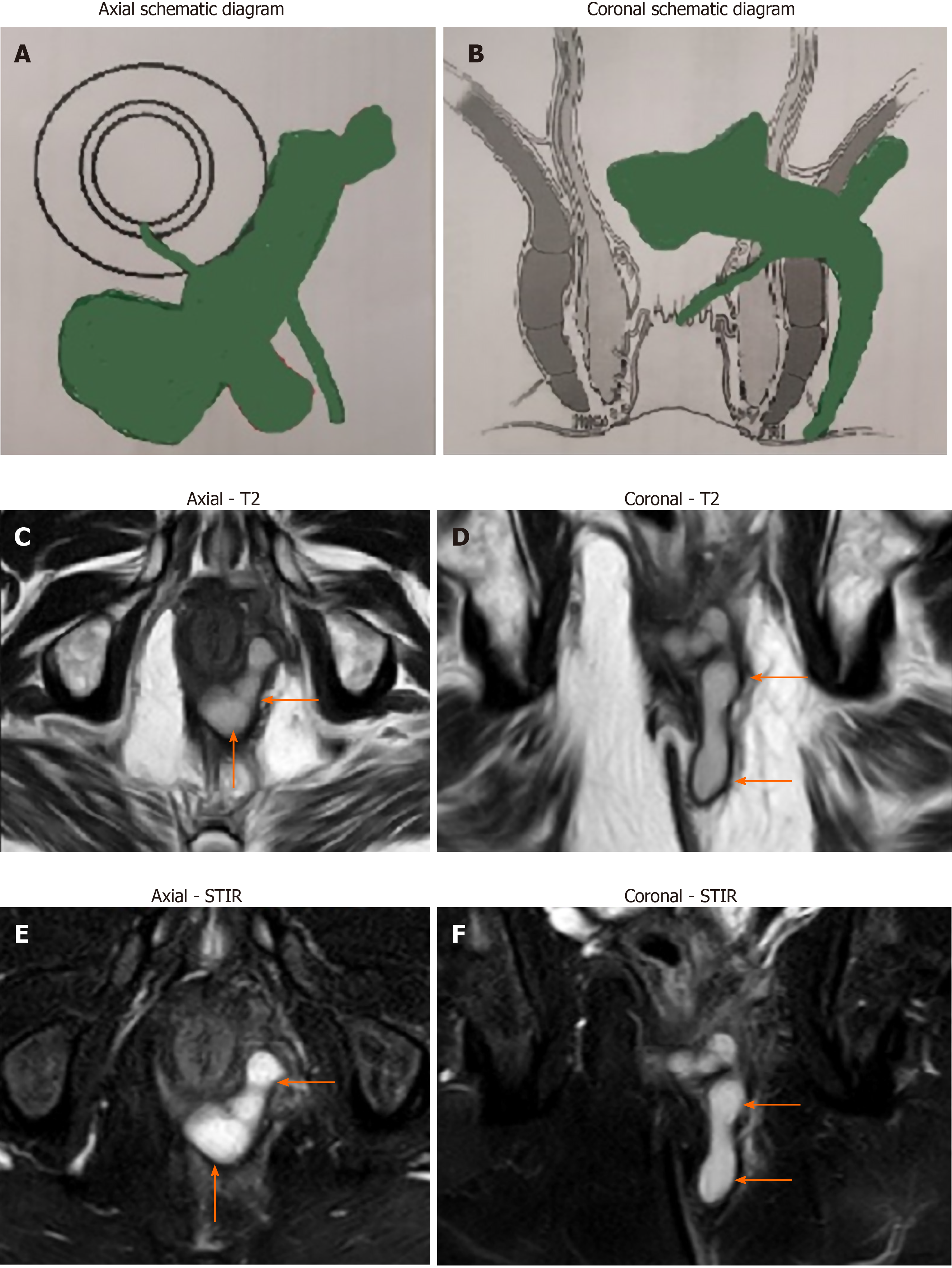
Figure 1 A 68-year-old male patient with tuberculosis infected high transsphincteric horseshoe complex anal fistula with multiple tracts (orange arrows show fistula tracts).
A: Schematic diagram-axial section; B: Schematic diagram-coronal section; C: magnetic resonance imaging (MRI)-axial section-T2 sequence; D: MRI-coronal section-T2 sequence; E: MRI-axial section-short T-1 inversion recovery sequence; and F: MRI-coronal section-short T-1 inversion recovery sequence.
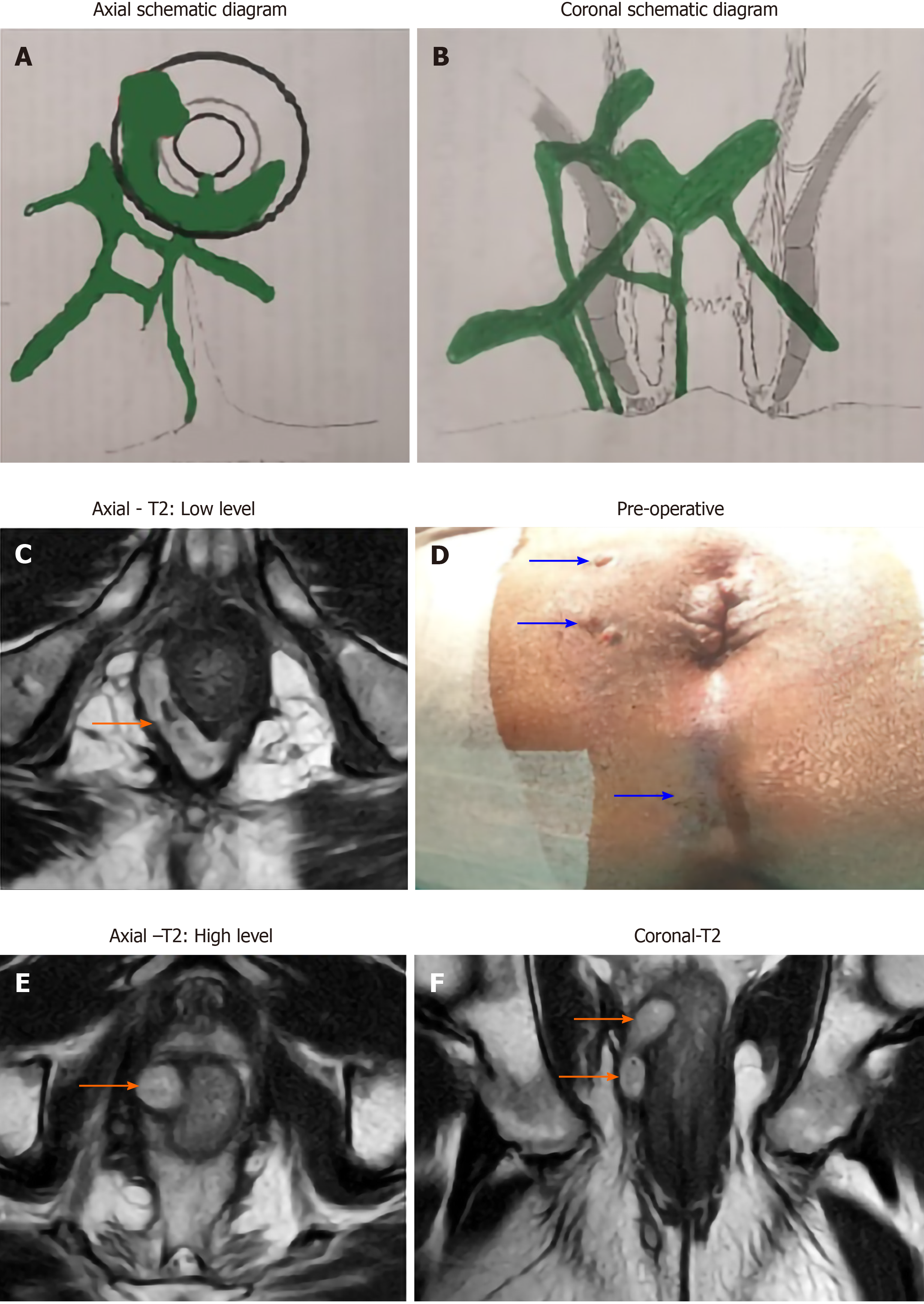
Figure 2 A 52-year-old male patient with tuberculosis infected high supralevator horseshoe complex anal fistula associated with an abscess and multiple tracts.
There is a supralevator rectal opening at 10 o'clock (orange arrows show fistula tracts). A: Schematic diagram-axial section; B: Schematic diagram-coronal section; C: Magnetic resonance imaging (MRI)-axial section-T2 sequence-low level; D: Preoperative photograph showing multiple (three) external openings in perianal skin (marked by blue arrows); E: MRI-axial section-T2 sequence-high level showing a supralevator rectal opening at 10 o'clock; and F: MRI-coronal section-T2 sequence.
- Citation: Garg P, Goyal A, Yagnik VD, Dawka S, Menon GR. Diagnosis of anorectal tuberculosis by polymerase chain reaction, GeneXpert and histopathology in 1336 samples in 776 anal fistula patients. World J Gastrointest Surg 2021; 13(4): 355-365
- URL: https://www.wjgnet.com/1948-9366/full/v13/i4/355.htm
- DOI: https://dx.doi.org/10.4240/wjgs.v13.i4.355


