Copyright
©The Author(s) 2021.
World J Gastrointest Surg. Mar 27, 2021; 13(3): 315-322
Published online Mar 27, 2021. doi: 10.4240/wjgs.v13.i3.315
Published online Mar 27, 2021. doi: 10.4240/wjgs.v13.i3.315
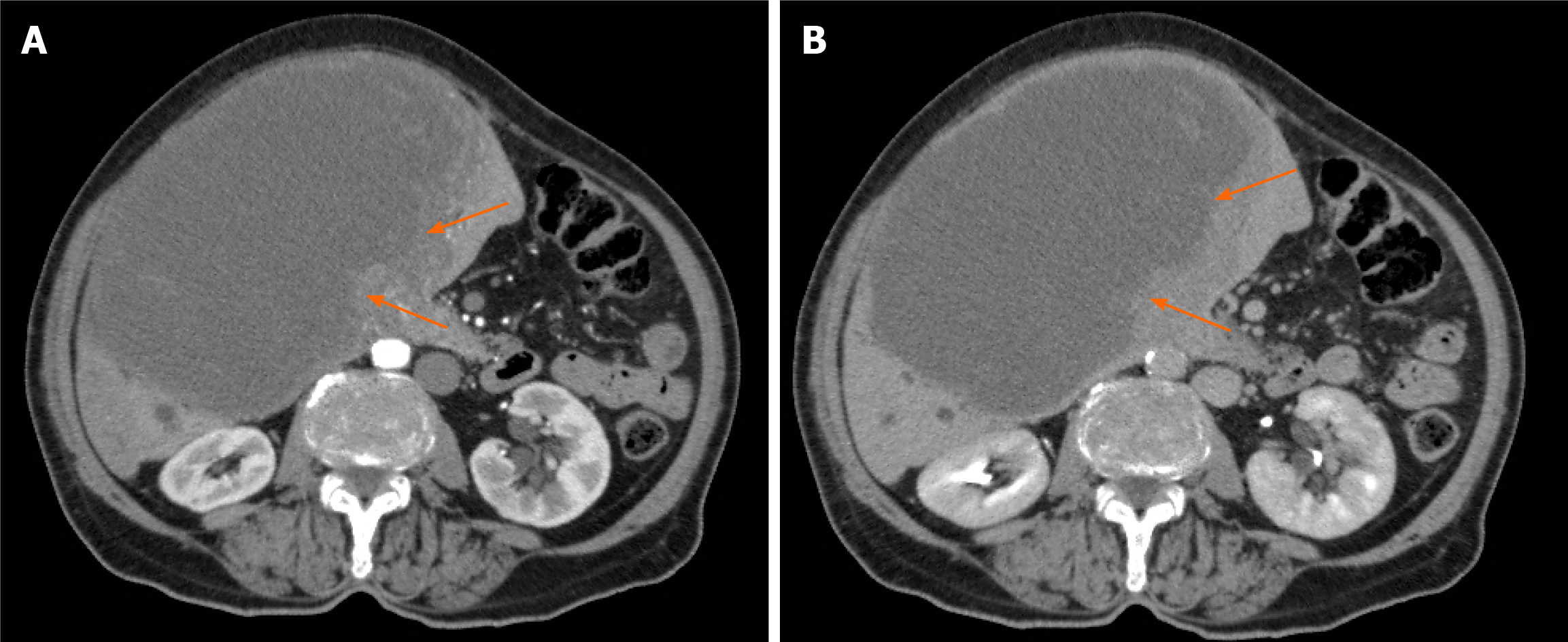
Figure 1 Abdominal computed tomography showing an expansive hepatic mass with a solid component (arrows).
A: Intense arterial enhancement; B: Late-phase washout were present.
Figure 2 Magnetic resonance imaging showing a large, central, T1-hyperintense hepatic lesion.
A: Blood products (asterisk); B: Arterial enhancement of the solid component (arrow); C: Magnetic resonance cholangiogram showing dilation of the intrahepatic bile ducts.
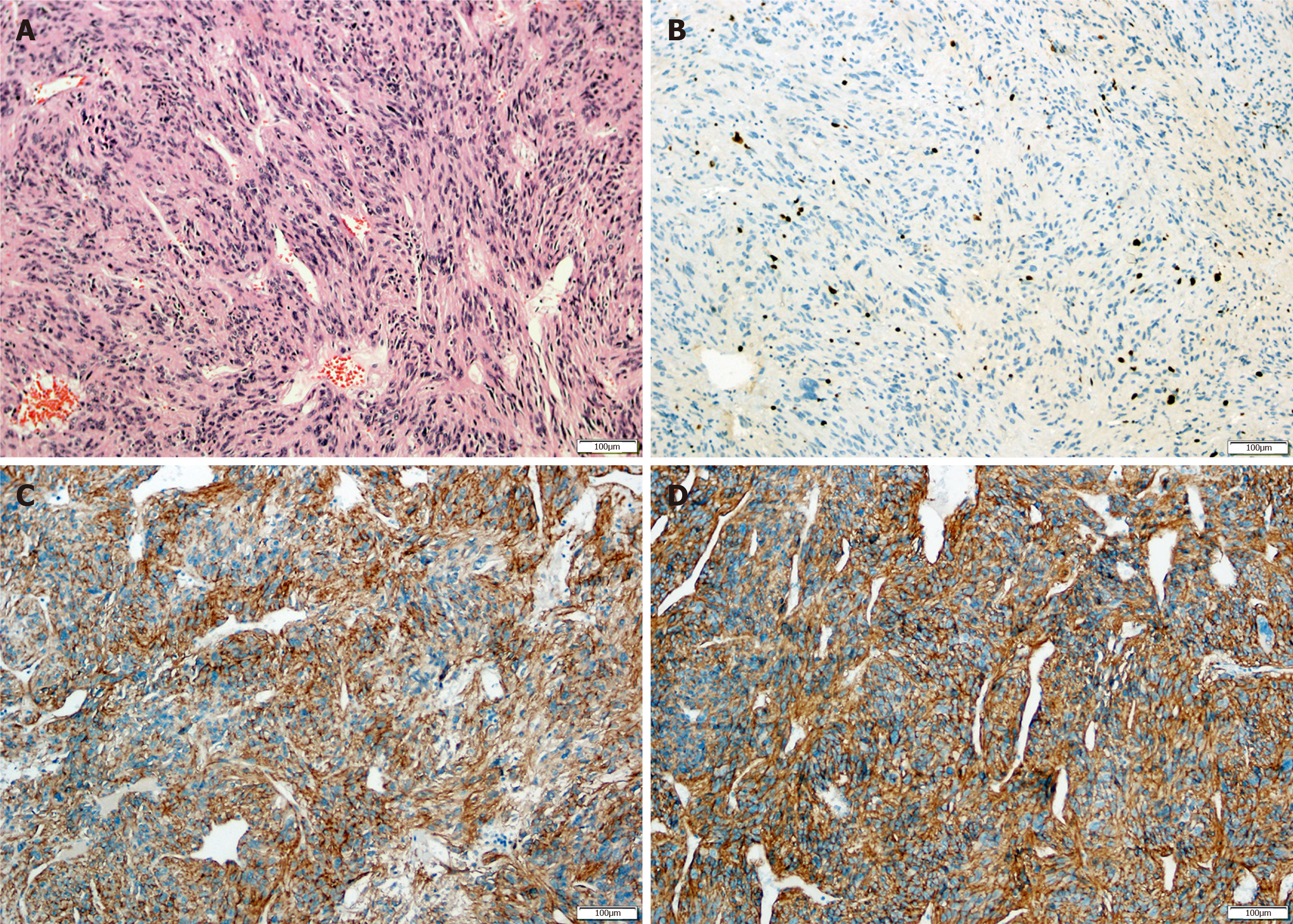
Figure 3 Histopathological examination.
A: bundles of spindle-shaped cells in an irregular pattern, eosinophilic cytoplasm, with normal hepatic parenchyma in peripheral areas in hematoxylin-eosin staining (magnification, × 100); B-D: Immunohistochemical analysis showed that the tumor was positive for Ki-67 (B), CD-117 (C) and DOG1 (D) (magnification, × 100).
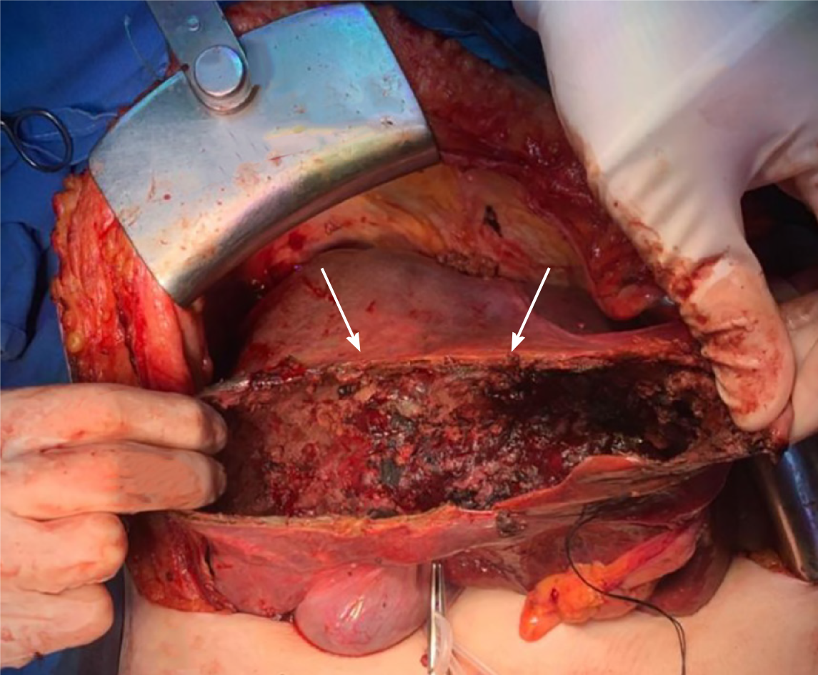
Figure 4 Area of tumor resection.
Arrows show the residual tumor capsule.
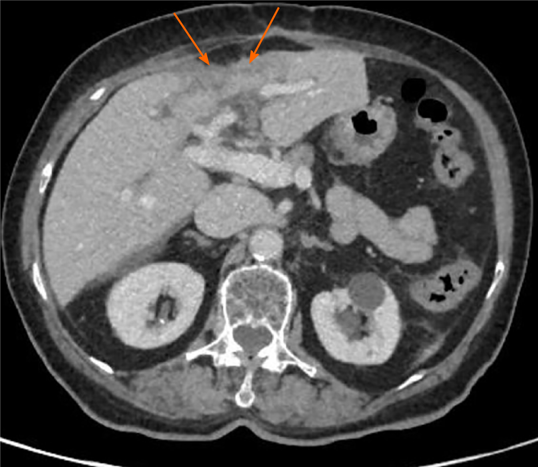
Figure 5 Follow-up abdominal computed tomography scan showing significant reduction of the hepatic mass (arrows), its solid component, and the mass effect on adjacent structures.
- Citation: Fernandes MR, Ghezzi CLA, Grezzana-Filho TJ, Feier FH, Leipnitz I, Chedid AD, Cerski CTS, Chedid MF, Kruel CRP. Giant hepatic extra-gastrointestinal stromal tumor treated with cytoreductive surgery and adjuvant systemic therapy: A case report and review of literature. World J Gastrointest Surg 2021; 13(3): 315-322
- URL: https://www.wjgnet.com/1948-9366/full/v13/i3/315.htm
- DOI: https://dx.doi.org/10.4240/wjgs.v13.i3.315


