Copyright
©The Author(s) 2021.
World J Gastrointest Surg. Dec 27, 2021; 13(12): 1628-1637
Published online Dec 27, 2021. doi: 10.4240/wjgs.v13.i12.1628
Published online Dec 27, 2021. doi: 10.4240/wjgs.v13.i12.1628
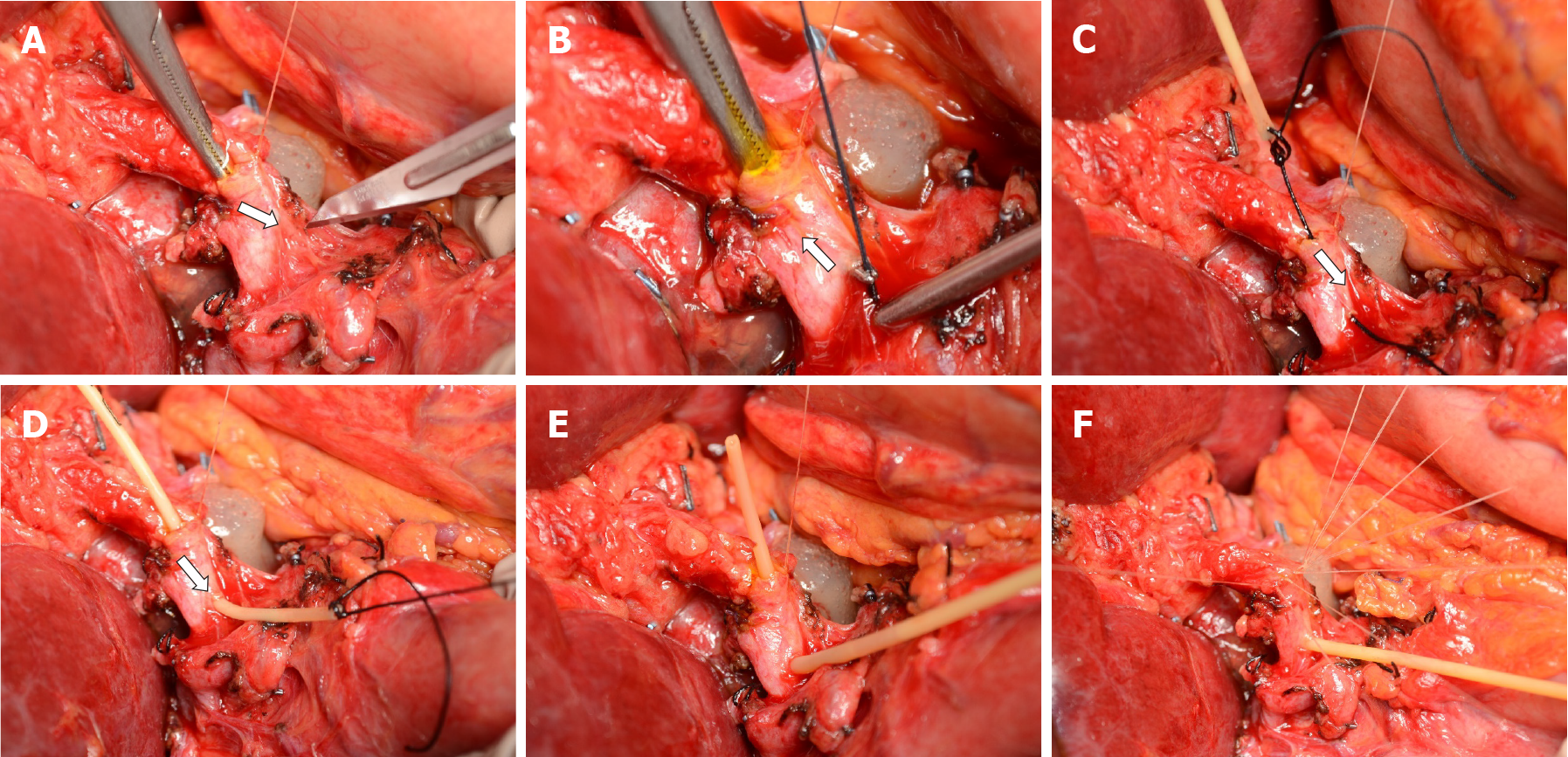
Figure 1 T-tube insertion protocol.
A: The right-angle is advanced through the open anterior layer of the duct-to-duct anastomosis and a choledochotomy is created with a no. 11 scalpel; B-D: A silk tie is grabbed and pulled through the choledochotomy after being stitched to the horizontal end of the T-tube; E: the T-tube is allocated inside the bile duct; F: the anastomosis is completed with interrupted Vicryl 6/0 stitches.
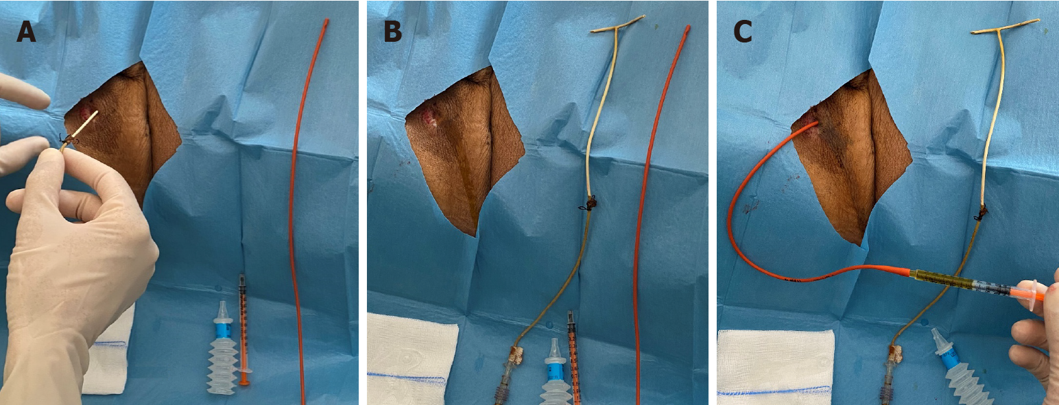
Figure 2 Bedside, standard T-tube removal procedure.
A: The T-tube is removed; B: A Nelaton drain is kept aside to measure the length of the T-tube internal tract (whiter portion of the T-tube); C: The Nelaton drain is inserted approximately 2 cm shorter than the measured length.
- Citation: Spoletini G, Bianco G, Franco A, Frongillo F, Nure E, Giovinazzo F, Galiandro F, Tringali A, Perri V, Costamagna G, Avolio AW, Agnes S. Pediatric T-tube in adult liver transplantation: Technical refinements of insertion and removal. World J Gastrointest Surg 2021; 13(12): 1628-1637
- URL: https://www.wjgnet.com/1948-9366/full/v13/i12/1628.htm
- DOI: https://dx.doi.org/10.4240/wjgs.v13.i12.1628


