Copyright
©The Author(s) 2021.
World J Gastrointest Surg. Nov 27, 2021; 13(11): 1327-1337
Published online Nov 27, 2021. doi: 10.4240/wjgs.v13.i11.1327
Published online Nov 27, 2021. doi: 10.4240/wjgs.v13.i11.1327
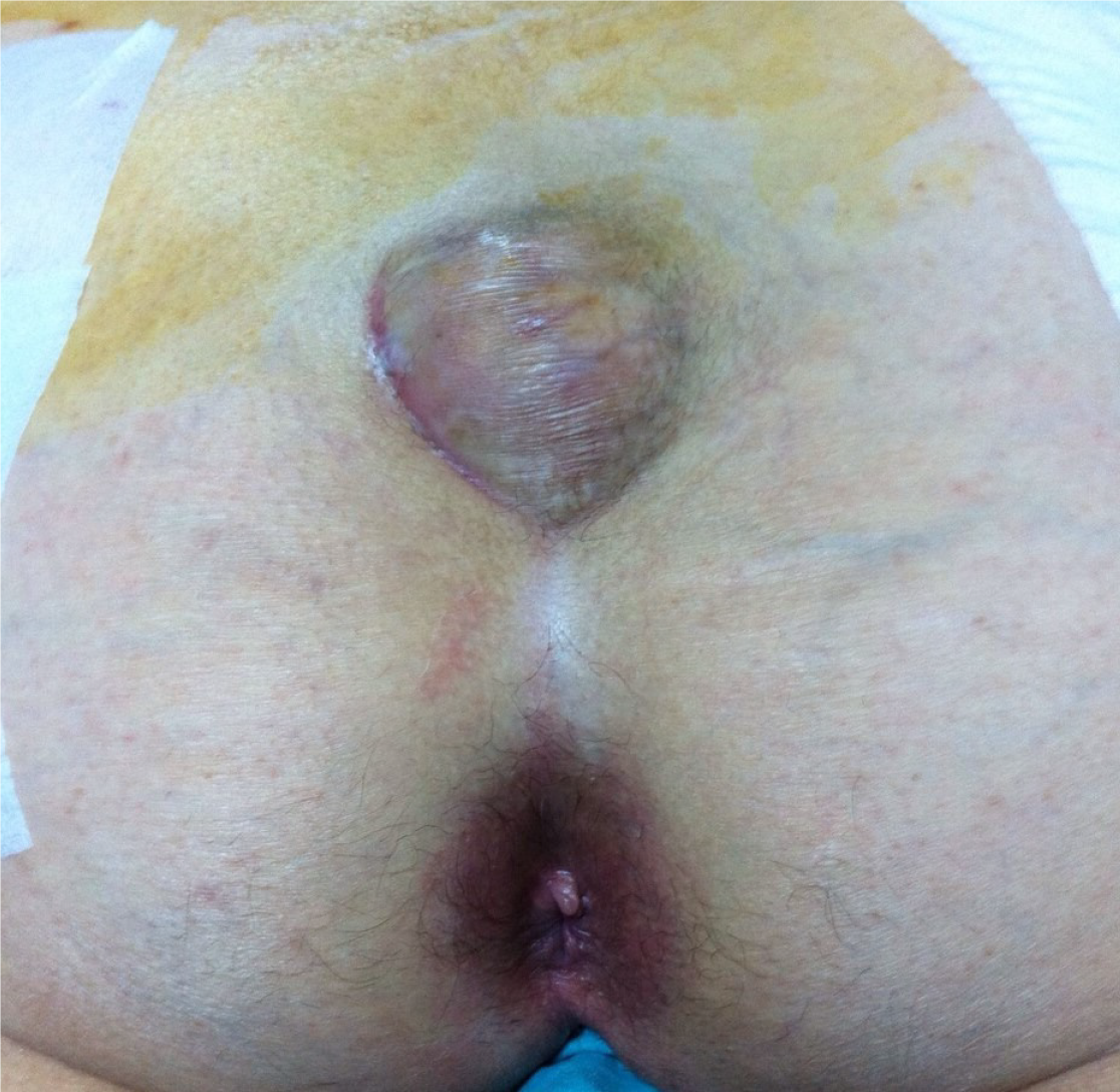
Figure 1
A patient presented with complaints of recurrent fistula, which was ultimately diagnosed as epidermoid cyst.
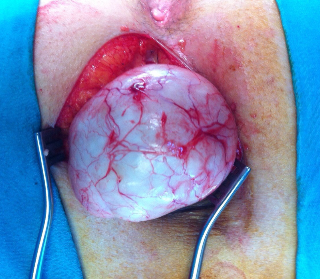
Figure 2
Intraoperative image of the epidermoid cyst.
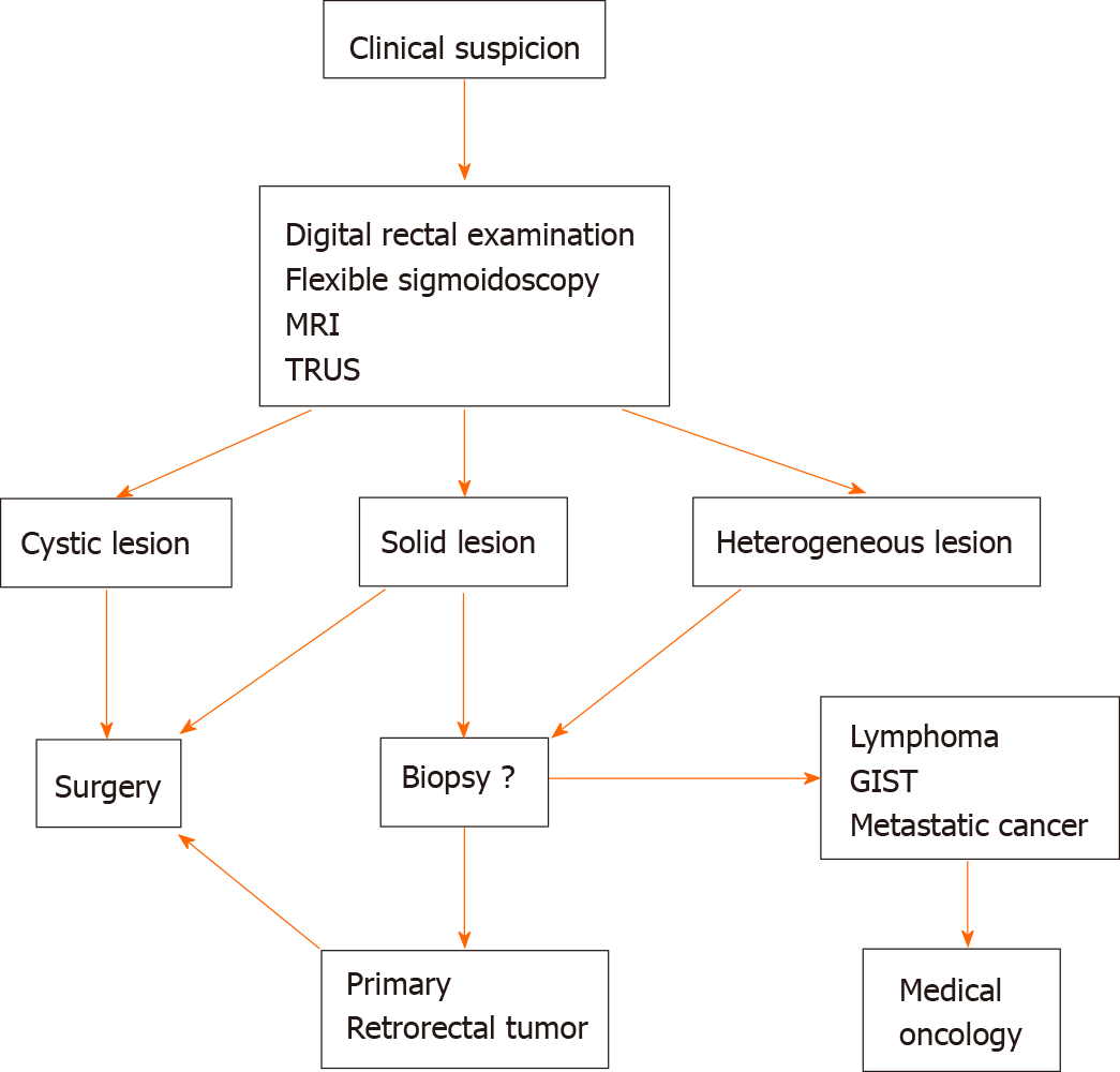
Figure 3 Flow diagram for the management of retrorectal tumors.
MRI: Magnetic resonance imaging; GIST: Gastrointestinal stromal tumor; TRUS: Transrectal ultrasonography.
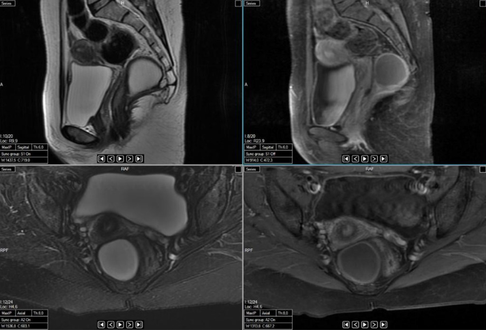
Figure 4
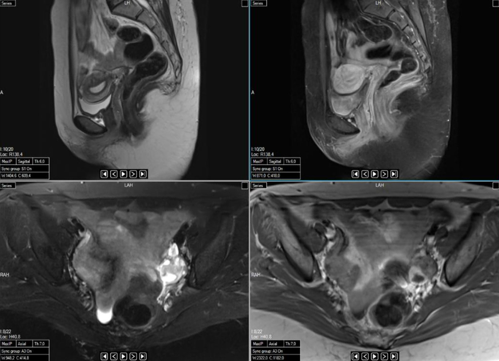
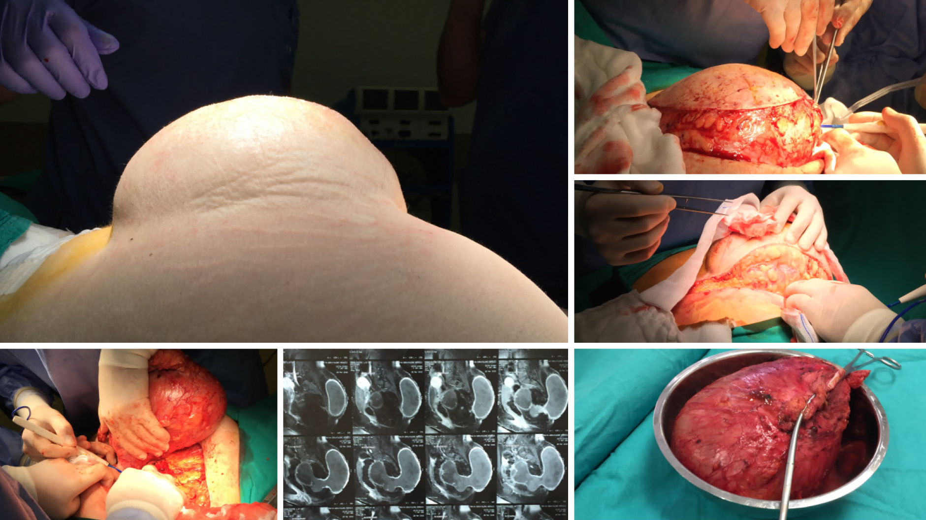
Sagittal and axial magnetic resonance images showing a cystic teratoma localized in the retrorectal area.
Figure 5
Sagittal and axial magnetic resonance images taken after resectioning the cystic teratoma shown in Figure 4.
Figure 6
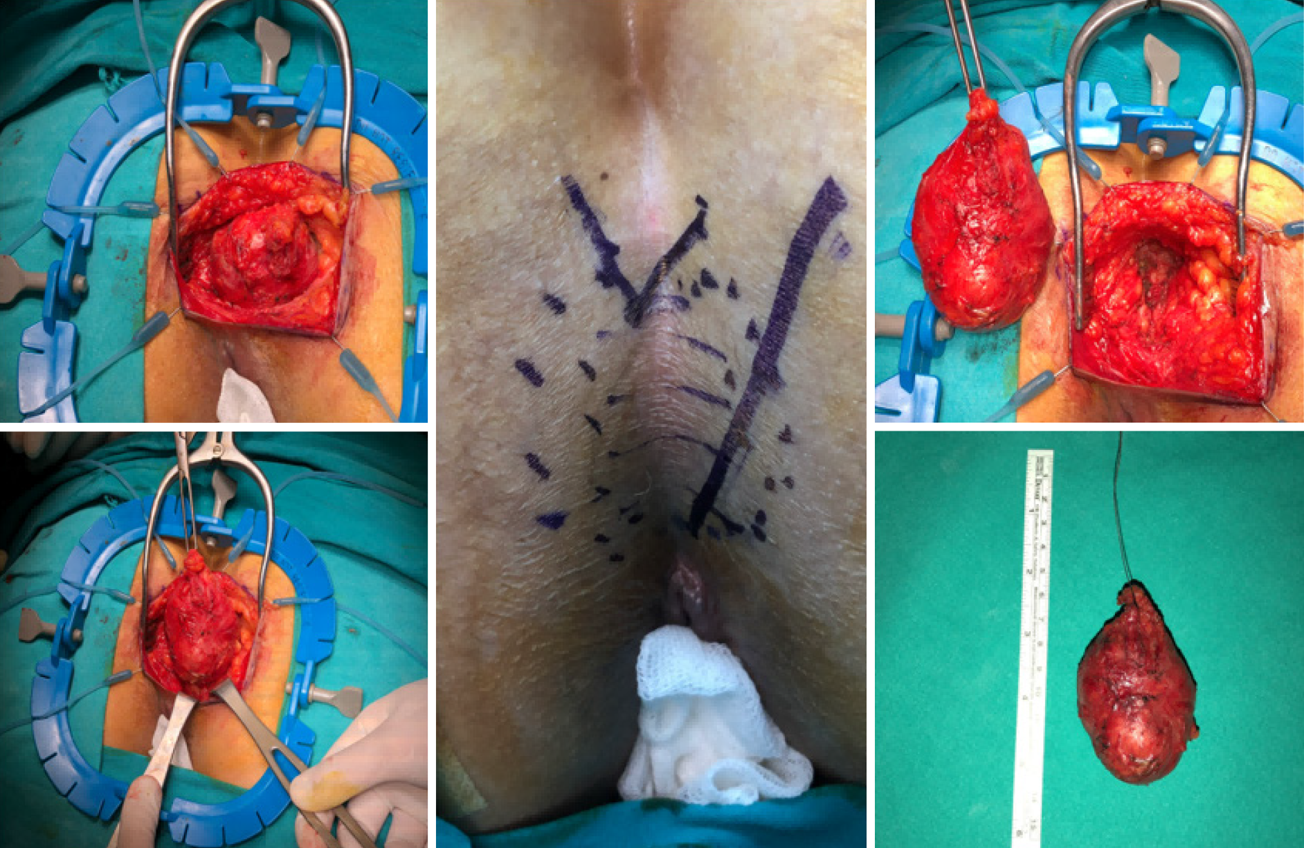
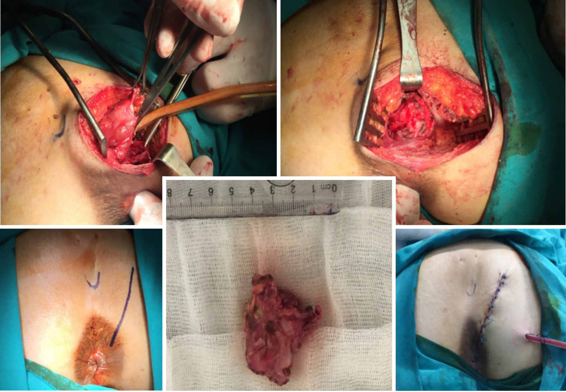
Perineal approach via parasagittal incision in a patient with a tail-gut cyst.
Figure 7
Perineal approach via parasagittal incision in a patient with an epidermoid cyst.
Figure 8
Perineal approach via parasagittal incision in a patient with a teratoma.
- Citation: Balci B, Yildiz A, Leventoğlu S, Mentes B. Retrorectal tumors: A challenge for the surgeons. World J Gastrointest Surg 2021; 13(11): 1327-1337
- URL: https://www.wjgnet.com/1948-9366/full/v13/i11/1327.htm
- DOI: https://dx.doi.org/10.4240/wjgs.v13.i11.1327


