Copyright
©The Author(s) 2021.
World J Gastrointest Surg. Oct 27, 2021; 13(10): 1190-1201
Published online Oct 27, 2021. doi: 10.4240/wjgs.v13.i10.1190
Published online Oct 27, 2021. doi: 10.4240/wjgs.v13.i10.1190
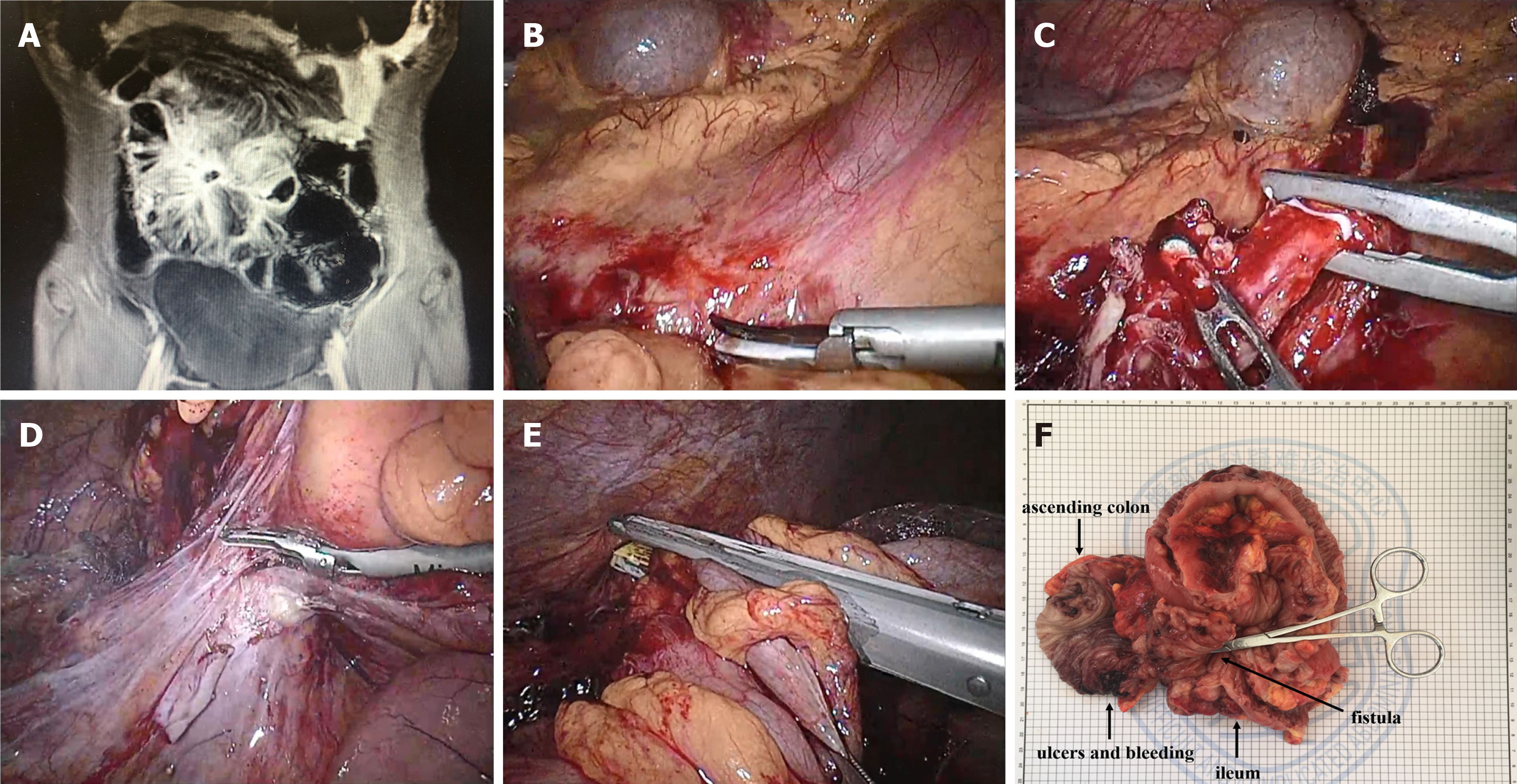
Figure 1 Laparoscopic ileocecal resection.
A: Magnetic resonance enterography showed that the ileum was adhered into a mass with the possible formation of partial enteral fistula; B: Separation of the superior mesenteric vein; C: Isolation of the ileal blood vessels; D: Separation of the ileocecum; E: Disconnection of the small intestine; F: Specimen of the diseased bowel.
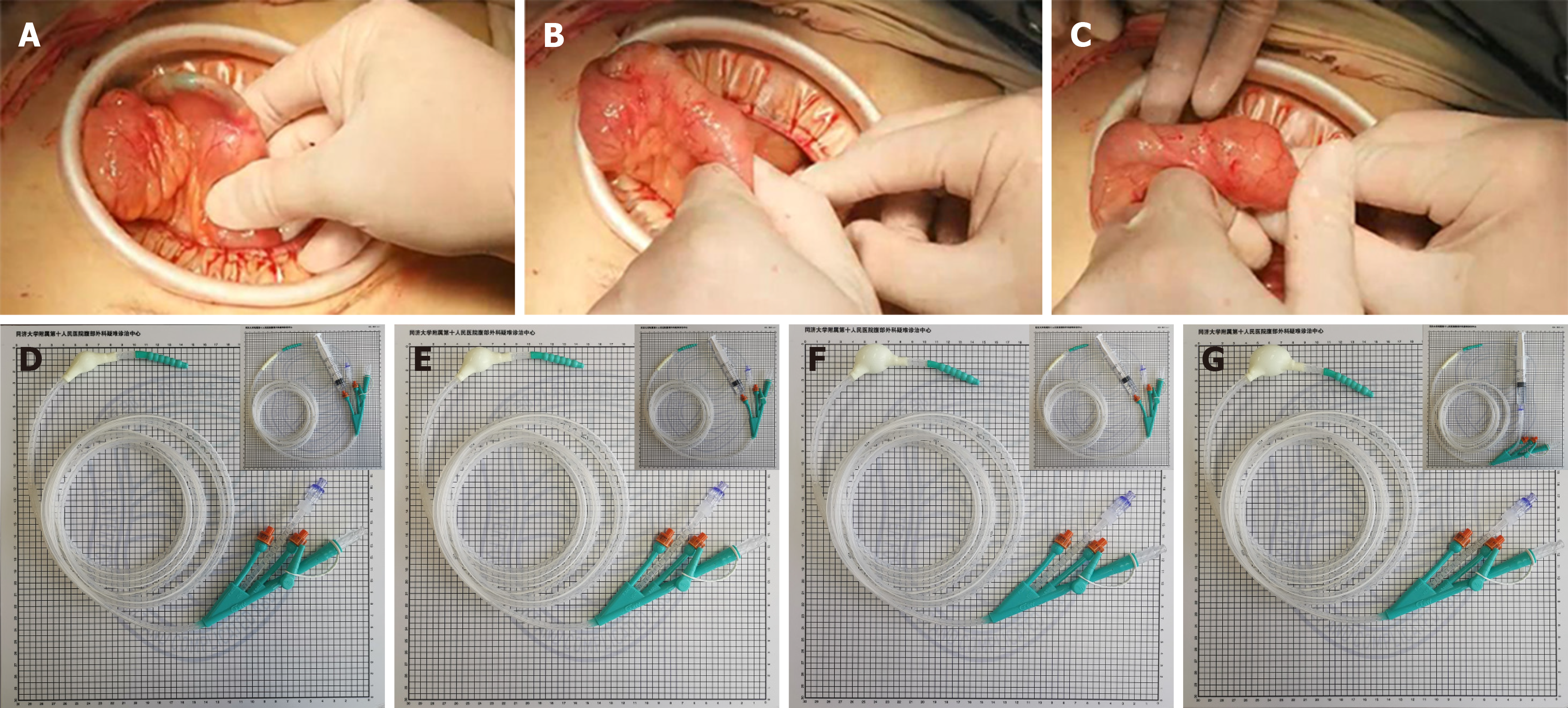
Figure 2 Intraoperative balloon dilatation and different balloon sizes of the ileus tube.
A: Balloon prior to segment of enterostenosis; B: Balloon passes through segment of enterostenosis; C: Balloon posterior to segment of enterostenosis; D: A balloon with a diameter of 1.5 cm can be formed by injecting 3 mL of sterilized water; E: A balloon with a diameter of 2 cm can be formed by injecting 5 mL of sterilized water; F: A balloon with a diameter of 2.5 cm can be formed by injecting 8 mL of sterilized water; G: A balloon with a diameter of 3 cm can be formed by injecting 10 mL of sterilized water.
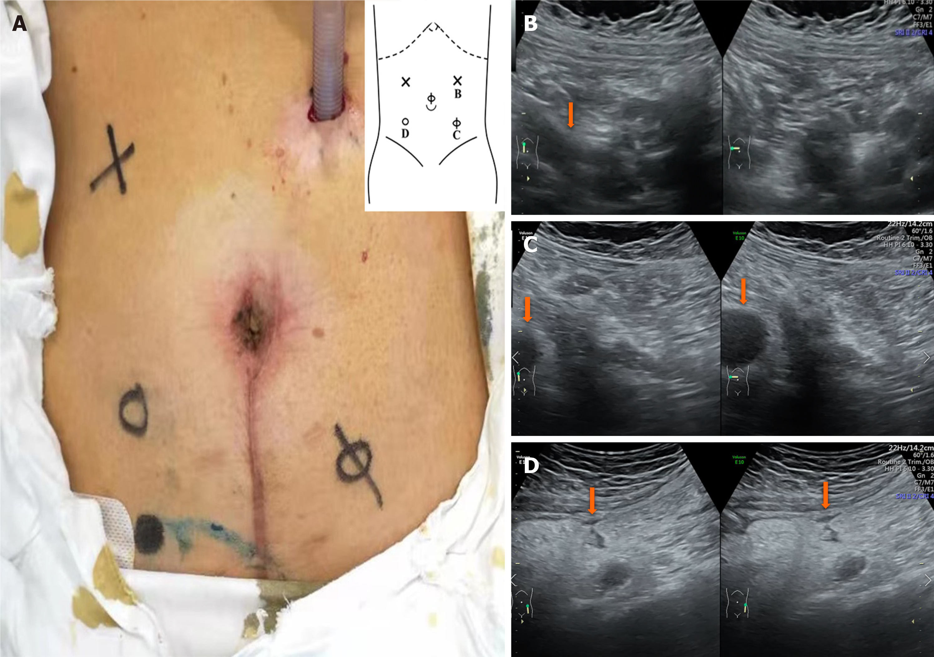
Figure 3 With the changes during respiration, B-ultrasonography was used to examine different areas to determine the activity between the abdominal wall and viscera.
A: The degree of intraperitoneal adhesion is indicated by the annotations "×", "Φ", and "loop". "×" means that the activity is greater than 30 mm, the activity is better in the abdominal cavity, and no adhesion is considered. "Φ" means that the activity is greater than 5 mm, but less than 30 mm, and moderate intraperitoneal activity and moderate adhesion are considered. "loop" means that the activity is less than 5 mm, and severe adhesion is considered; B: In (A), the area denoted "×" in the left upper abdomen is seen under ultrasound. The blood vessels disappear from the screen, as shown in the arrow. Considering that there is no adhesion, it is the first place to puncture; C: In (A), the area denoted "Φ" in the right lower abdomen is seen under ultrasound. The range of intraperitoneal vessels with respiration is shown by the arrow; D: In (A), the area denoted "loop" in the right lower abdomen is seen under ultrasound. The range of intraperitoneal vessels with respiration is shown by the arrow.
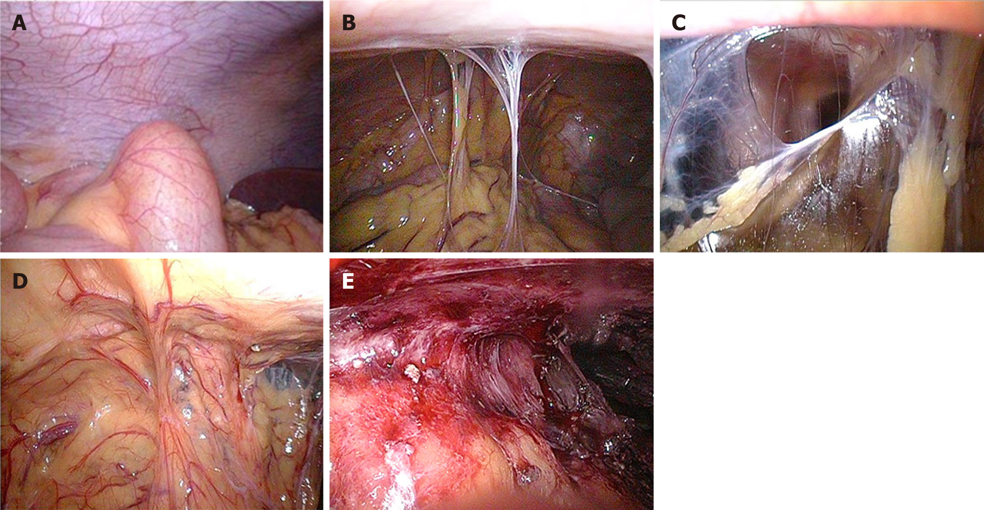
Figure 4 Levels of abdominal adhesion.
A: Level 0, no adhesion; B: Level 1, slight adhesion (strip adhesion); C: Level 2, moderate adhesion (membrane adhesion or tight adhesion); D: Level 3, severe adhesion (fusion adhesion or complex adhesion); E: Level 4, extremely severe adhesion (wide compound adhesion).
- Citation: Wan J, Liu C, Yuan XQ, Yang MQ, Wu XC, Gao RY, Yin L, Chen CQ. Laparoscopy for Crohn's disease: A comprehensive exploration of minimally invasive surgical techniques. World J Gastrointest Surg 2021; 13(10): 1190-1201
- URL: https://www.wjgnet.com/1948-9366/full/v13/i10/1190.htm
- DOI: https://dx.doi.org/10.4240/wjgs.v13.i10.1190


