Copyright
©The Author(s) 2021.
World J Gastrointest Surg. Oct 27, 2021; 13(10): 1180-1189
Published online Oct 27, 2021. doi: 10.4240/wjgs.v13.i10.1180
Published online Oct 27, 2021. doi: 10.4240/wjgs.v13.i10.1180
Figure 1
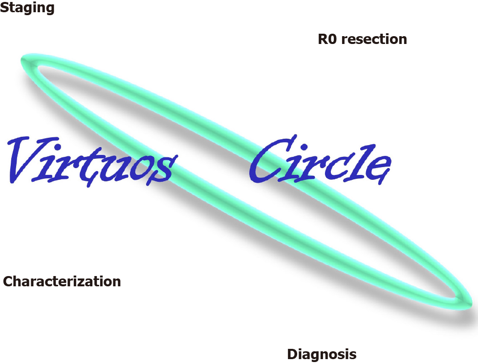
The “virtuous circle”: In a single step, it is possible to obtain R0-resection, characterization, diagnosis and histological staging of the lesion.
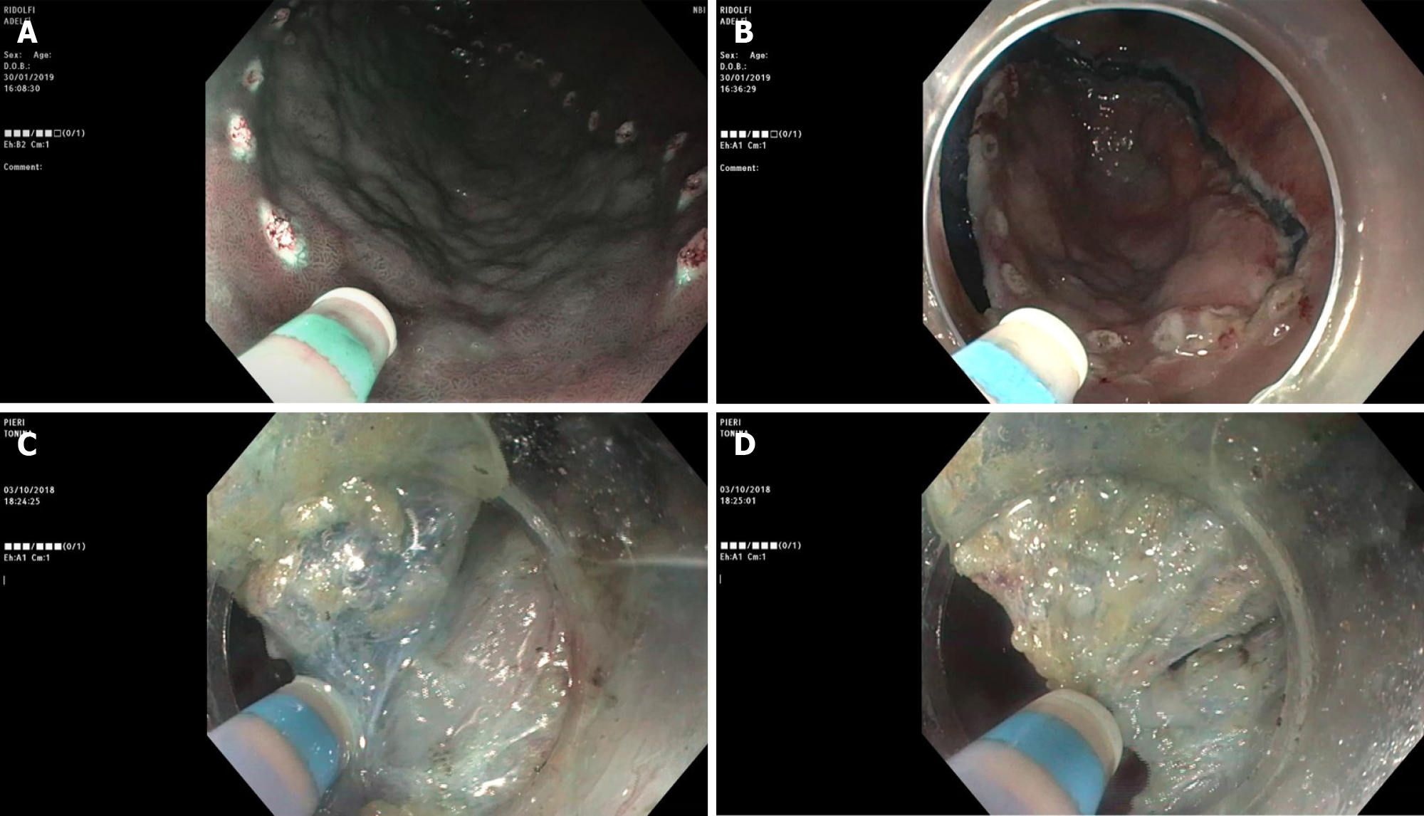
Figure 2 Multitasking device for multiple procedure steps.
A: Marking; B: Mucosal incision; C: Submucosal infiltration; D: Submucosal dissection.
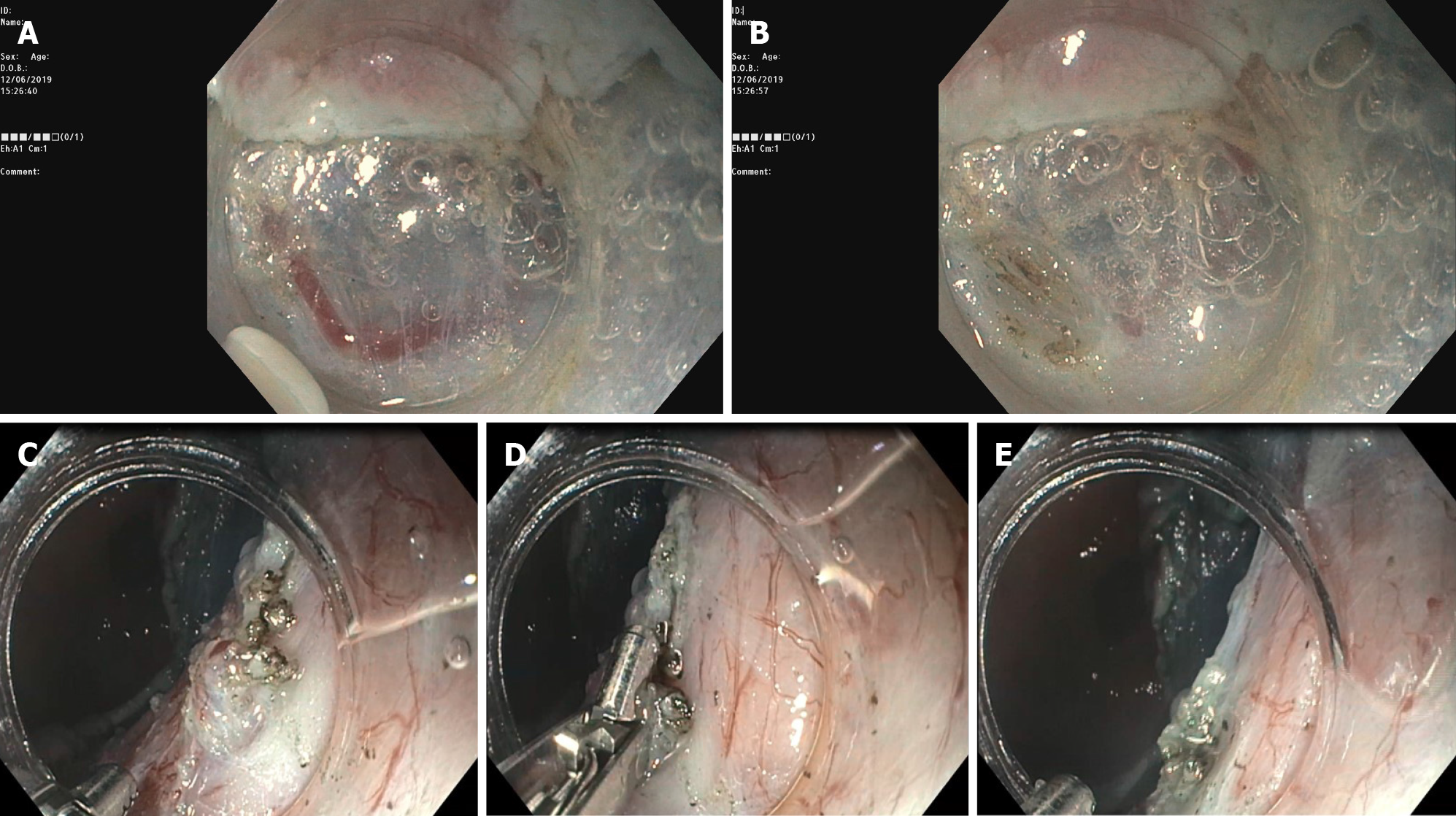
Figure 3 Hemostatic maneuver.
A: Submucosal visible vessel; B-E: Coagulation of the vessels with the endoscopic submucosal dissection knife or with forceps.
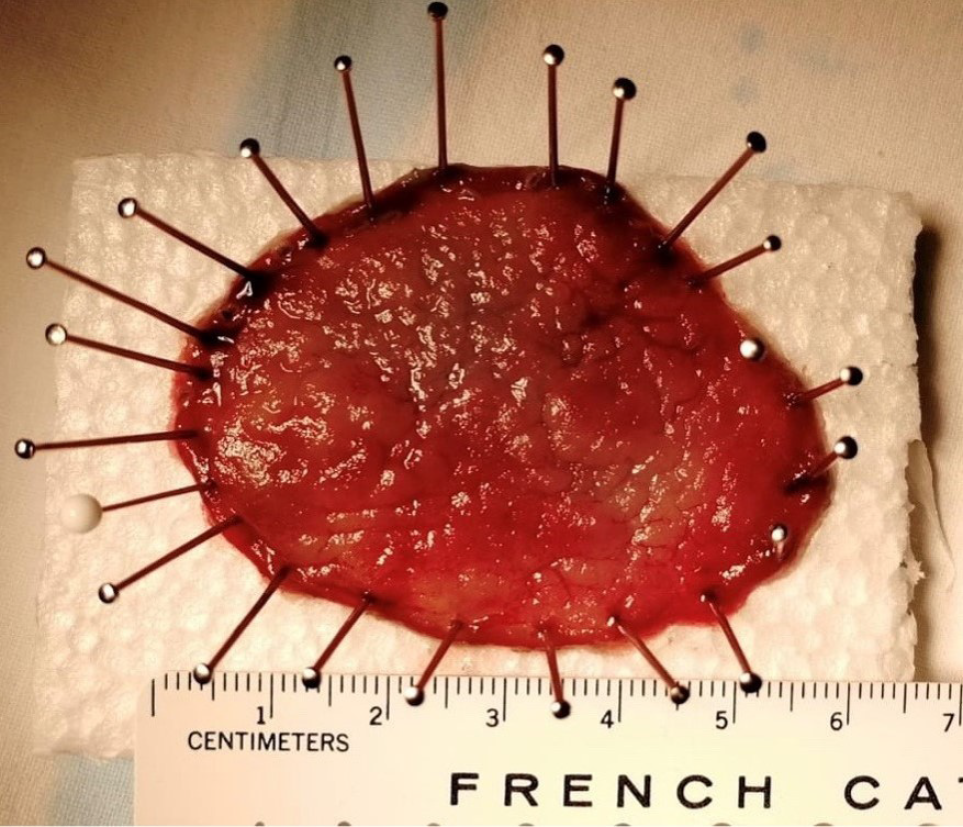
Figure 4
Whole resected specimen pinned down on a polystyrene block.
- Citation: De Luca L, Di Berardino M, Mangiavillano B, Repici A. Gastric endoscopic submucosal dissection in Western countries: Indications, applications, efficacy and training perspective. World J Gastrointest Surg 2021; 13(10): 1180-1189
- URL: https://www.wjgnet.com/1948-9366/full/v13/i10/1180.htm
- DOI: https://dx.doi.org/10.4240/wjgs.v13.i10.1180


