Copyright
©The Author(s) 2021.
World J Gastrointest Surg. Jan 27, 2021; 13(1): 30-49
Published online Jan 27, 2021. doi: 10.4240/wjgs.v13.i1.30
Published online Jan 27, 2021. doi: 10.4240/wjgs.v13.i1.30
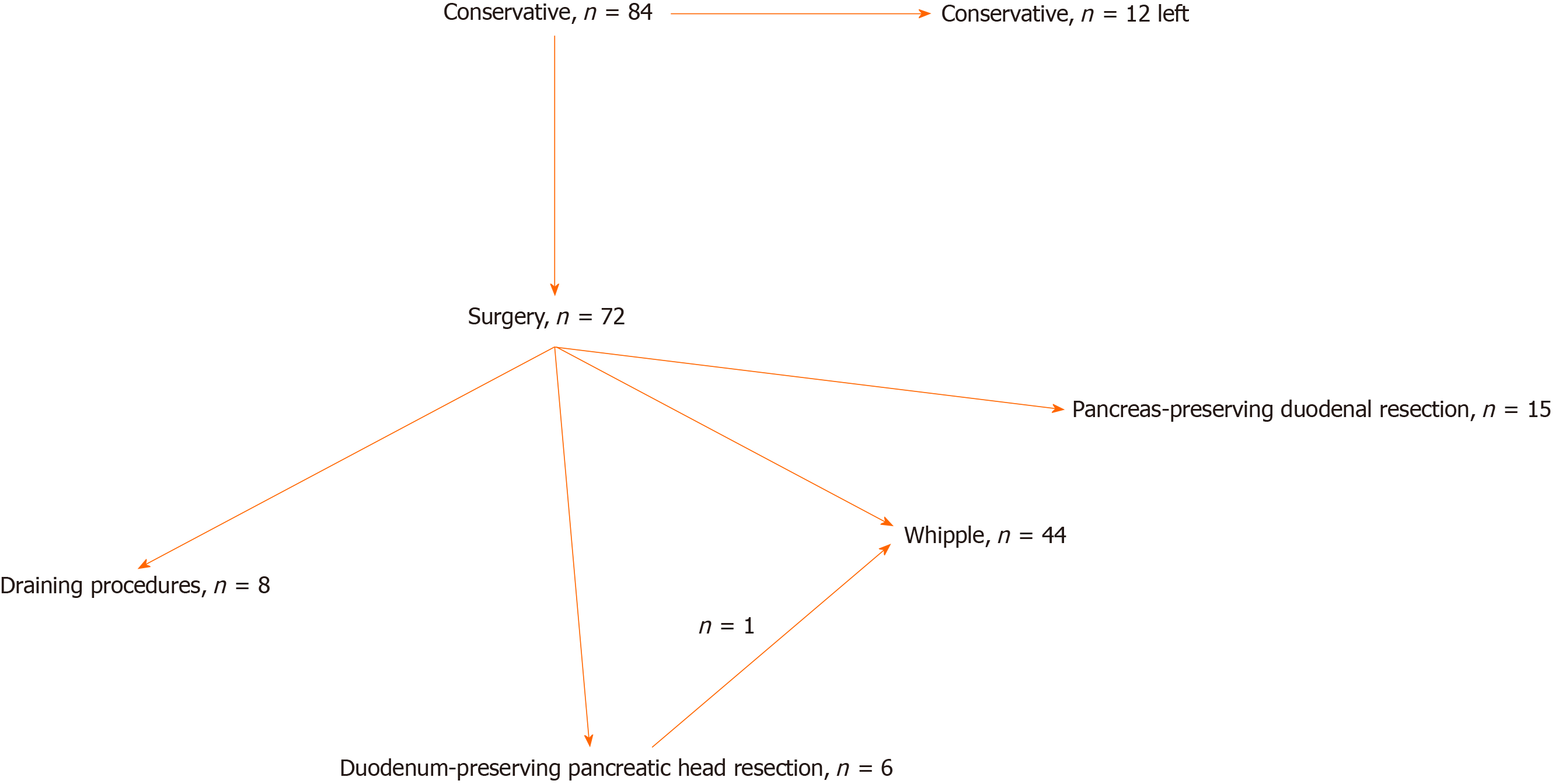
Figure 1 Patient flow chart.
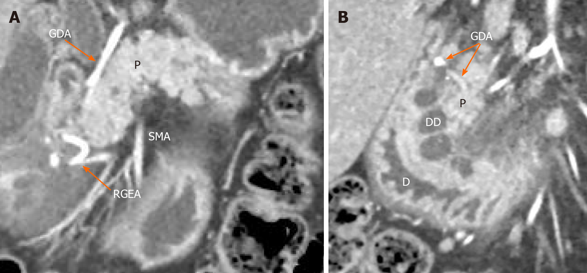
Figure 2 Isolated form of cystic dystrophy of the duodenal wall.
Arterial phase. Coronal view. A: Deformation and thickening of the medial wall of the duodenum (D), major papilla surrounded by well-defined cysts located in the submucosa (DD). The gastroduodenal artery is shifted forward and to the left, lying in the groove between the unaffected pancreatic head (P) and duodenal wall; B: Unchanged orthotopic pancreas. Only the duodenum and the groove are involved. SMA: Superior mesenteric artery; GDA: Gastroduodenal artery; RGEA: Right gastro-epiploic artery.
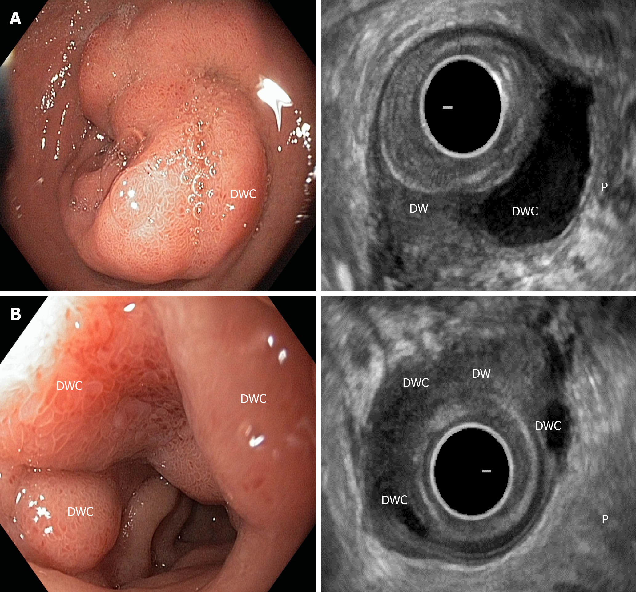
Figure 3 Duodenoscopy and endosonography.
Isolated form of the cystic dystrophy of the duodenal wall with unchanged orthotopic pancreas (P). A: Duodenal wall cyst (DWC) within the submucosa and muscularis of the diffusely thickened duodenal wall (DW); B: Multiple DWCs in the submucosa and muscularis surrounding the major papilla in the diffusely thickened DW. DWC: Duodenal wall cyst; DW: Duodenal wall.
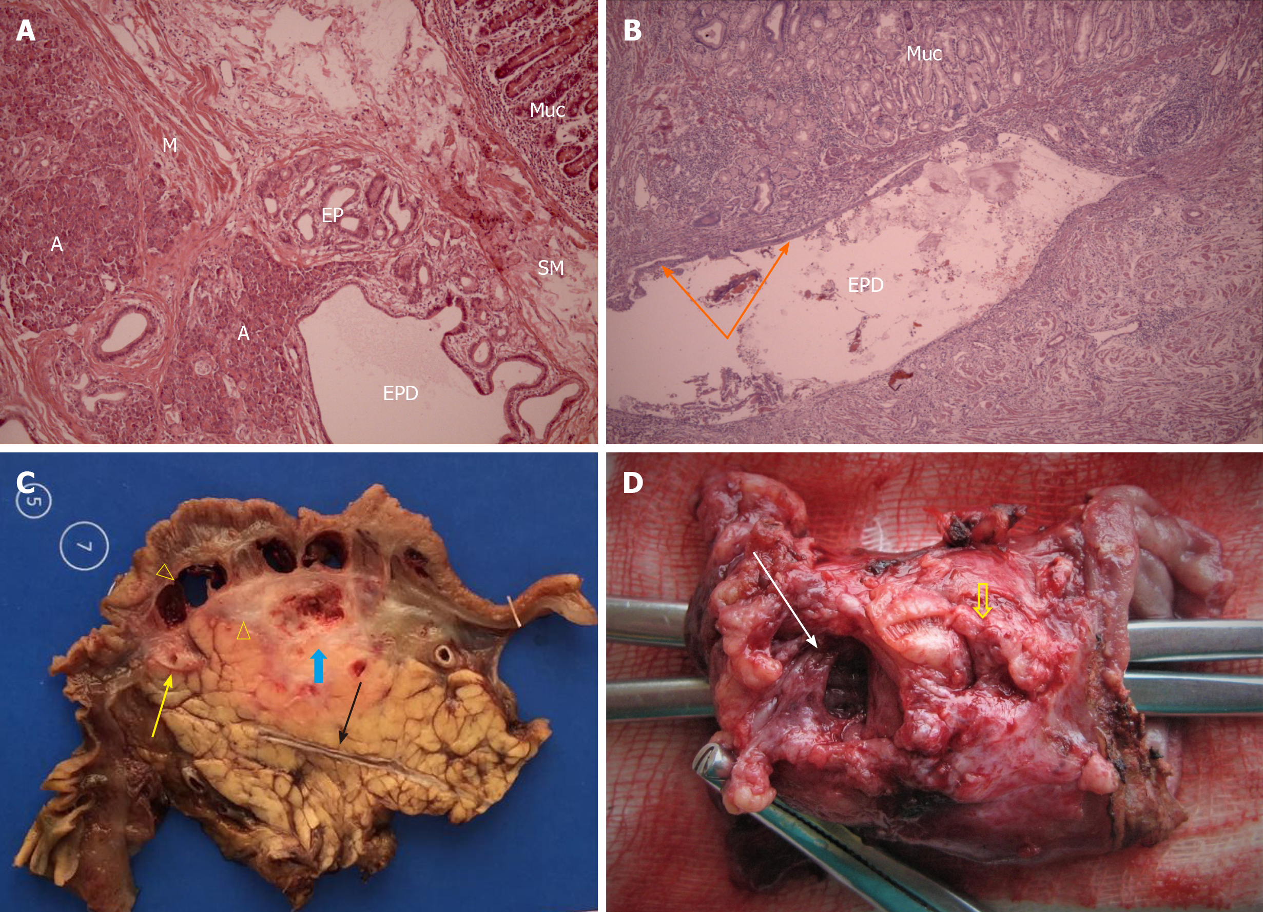
Figure 4 Microphotograph and resected specimen.
A: Microphotograph of the isolated form of the cystic dystrophy of the duodenal wall. Heterotopia of the pancreatic tissue in the duodenal wall. Ectopic pancreatic tissue (EP), dilated ducts of the ectopic pancreas (EPD) and acini (A) in the duodenal wall, M: Duodenal muscle layer fibers; SM: Duodenal submucosa; Muc: Duodenal mucosa. Hematoxylin-eosin, × 100; B: Microphotograph of the isolated form of the cystic dystrophy of the duodenal wall. Cyst in the duodenal wall formed by a dilated duct of the ectopic gland (EPD) with the foci of preserved epithelium (arrows). Hematoxylin-eosin, × 50; C: Cystic dystrophy of the duodenal wall associated with chronic pancreatitis in the pancreatic head (segmental form of groove pancreatitis). Resected specimen after Whipple procedure in a 47-year-old male. There are multiple cysts within the thickened, chronically inflamed duodenal wall of the second portion of the duodenum (arrowheads) without dilation of the common hepatic (yellow arrow) and main pancreatic (black arrow) ducts. Chronic inflammation in the pancreatic head with necrotic mass (thick blue arrow) makes pancreas-preserving surgery unjustified and pancreatoduodenectomy the surgery of choice; D: Isolated form of the cystic dystrophy of the duodenal wall = pure form of groove pancreatitis. Due to unchanged orthotopic gland, pancreas-preserving duodenal resection was performed in a 53-year-old male. Resected 6-cm specimen of the second part of the duodenum with major papilla (thick yellow arrow) and large scar-sided cyst of the medial duodenal wall with the remainder of the ectopic pancreatic tissue inside. A forceps was introduced into the duodenum to show the absence of communication between the duodenal lumen and the lumen of the cyst (white arrow).
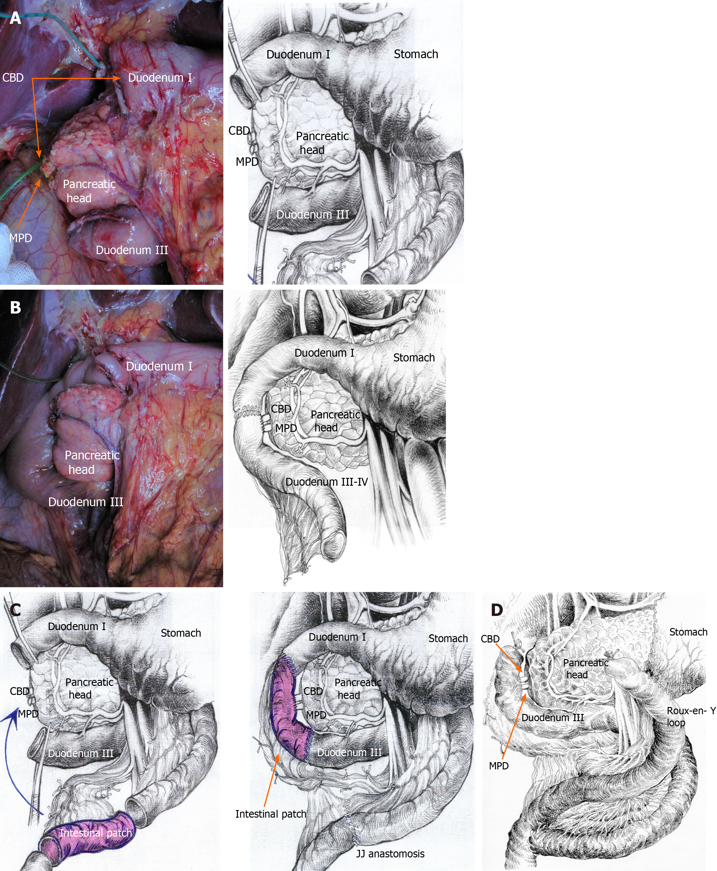
Figure 5 Isolated form of the cystic dystrophy of the duodenal wall.
Scheme of the pancreas-preserving resection of the second portion of the duodenum (A) with reconstruction by direct duodeno-jejunostomy (B), intestinal interposition (C) or Roux-en-Y method (D). CBD: Common bile duct; MPD; Main pancreatic duct.
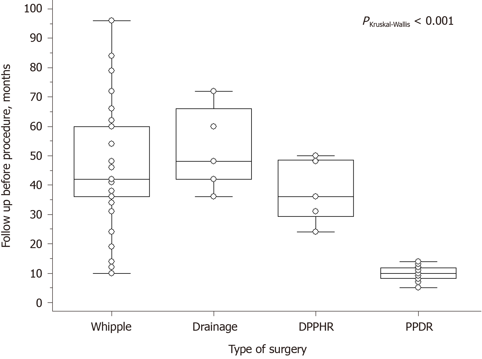
Figure 6 Duration of preoperative treatment of patients with cystic dystrophy of the duodenal wall.
Preoperative treatment before pancreas-preserving duodenal resection was significantly shorter when compared with the other subgroups (explanations in the text). PPDR: Pancreas-preserving duodenal resection; DPPHR: Duodenum-preserving pancreatic head resection.
- Citation: Egorov V, Petrov R, Schegolev A, Dubova E, Vankovich A, Kondratyev E, Dobriakov A, Kalinin D, Schvetz N, Poputchikova E. Pancreas-preserving duodenal resections vs pancreatoduodenectomy for groove pancreatitis. Should we revisit treatment algorithm for groove pancreatitis? World J Gastrointest Surg 2021; 13(1): 30-49
- URL: https://www.wjgnet.com/1948-9366/full/v13/i1/30.htm
- DOI: https://dx.doi.org/10.4240/wjgs.v13.i1.30


