Copyright
©The Author(s) 2020.
World J Gastrointest Surg. Aug 27, 2020; 12(8): 369-376
Published online Aug 27, 2020. doi: 10.4240/wjgs.v12.i8.369
Published online Aug 27, 2020. doi: 10.4240/wjgs.v12.i8.369
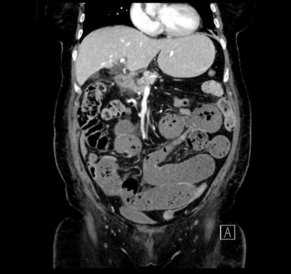
Figure 1 Computed tomography scan (coronal plane) showing small bowel dilatation and faecalisation.
The tip of the nasojejunal feeding tube was located appropriately within the efferent small bowel loop. There were also features of proximal to mid small bowel obstruction with faecalisation.
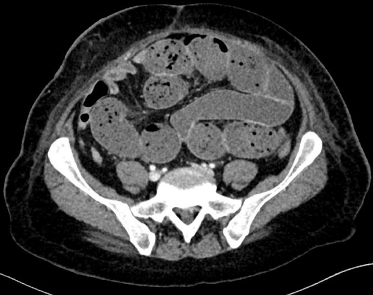
Figure 2 Computed tomography scan (axial plane) showing dilatation of the small bowel with faecalisation.
The transition point was not clearly defined Adhesions were postulated as a possible cause.
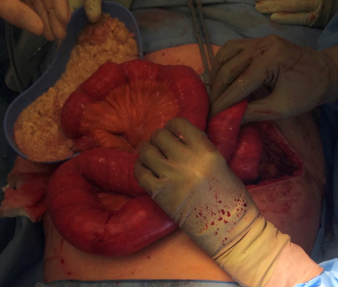
Figure 3 Intra-operative photography of intestinal decompression through an enterotomy, with evidence of enteral feed solidification.
These was full thickness, causing faecal and feed contamination of the peritoneum.
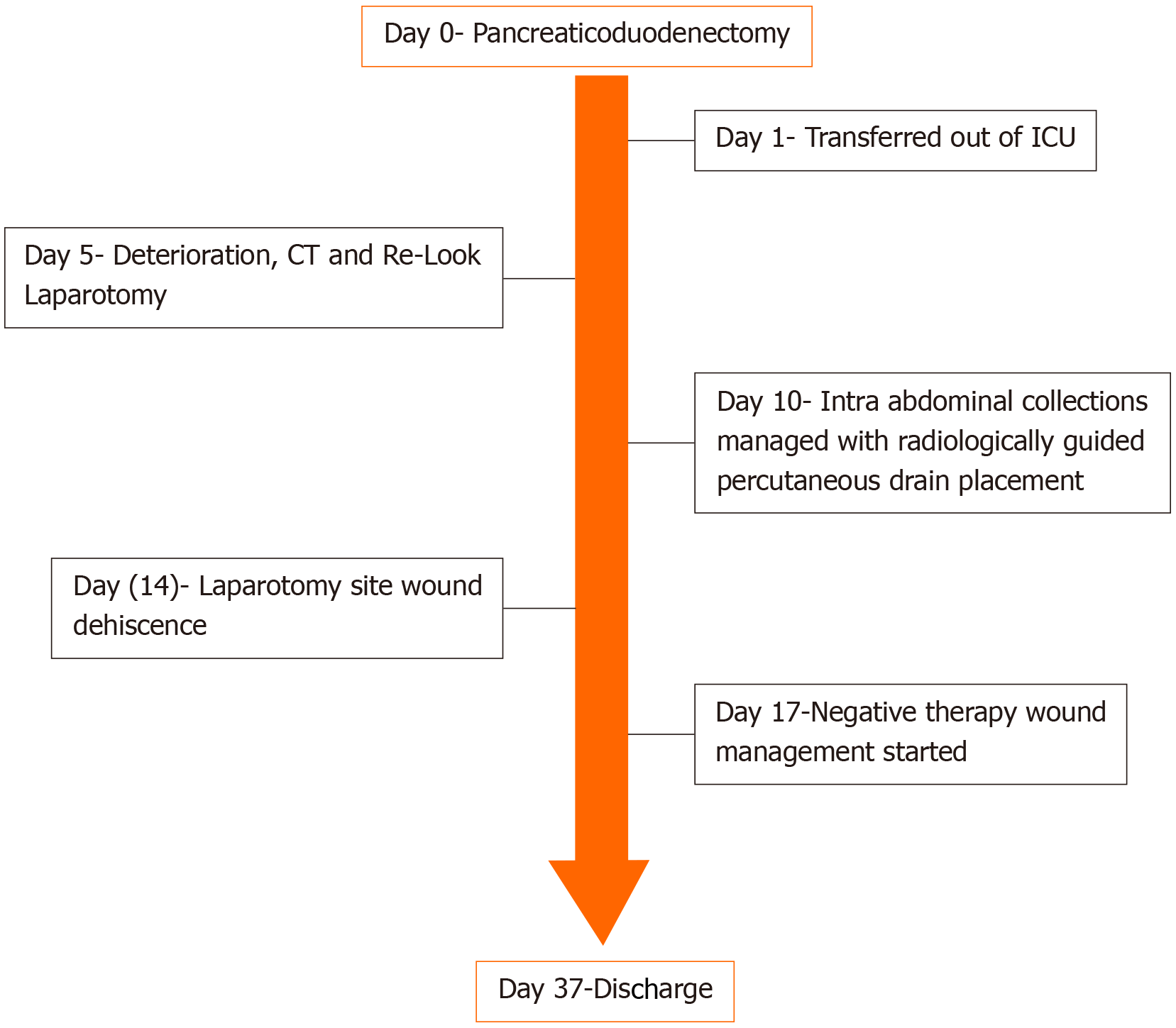
Figure 4 Timeline of case events.
ICU: Intensive care unit.
- Citation: Siddens ED, Al-Habbal Y, Bhandari M. Gastrointestinal obstruction secondary to enteral nutrition bezoar: A case report. World J Gastrointest Surg 2020; 12(8): 369-376
- URL: https://www.wjgnet.com/1948-9366/full/v12/i8/369.htm
- DOI: https://dx.doi.org/10.4240/wjgs.v12.i8.369


