Copyright
©The Author(s) 2020.
World J Gastrointest Surg. Jun 27, 2020; 12(6): 298-306
Published online Jun 27, 2020. doi: 10.4240/wjgs.v12.i6.298
Published online Jun 27, 2020. doi: 10.4240/wjgs.v12.i6.298
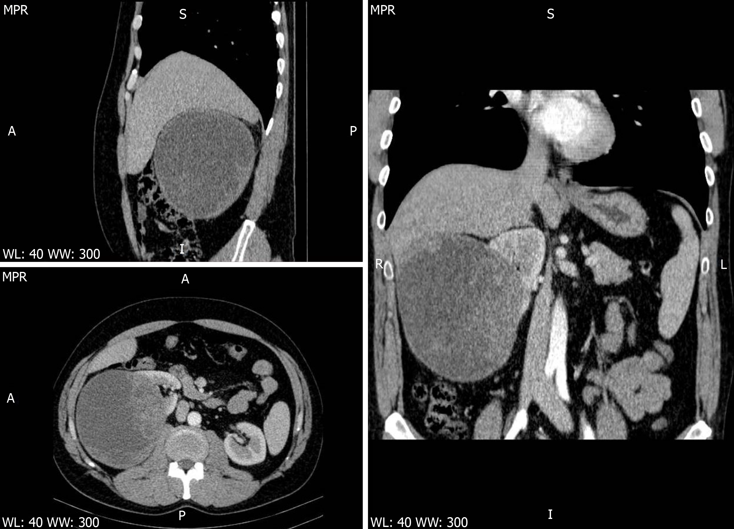
Figure 1 Computed tomography images.
Contrast-enhanced abdominal computed tomography scan thin multiplanar reconstruction, in nephrographic phase, shows a large well-defined predominantly cystic right renal mass, distorting the major calyces, with many solid enhancing intracystic areas, consistent with Bosniak type III renal cyst. No signs of right renal vein or inferior vena cava thrombosis (upper left-sagittal view, lower left-axial view, right-coronal view).
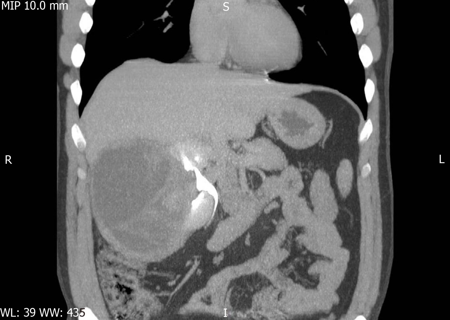
Figure 2 Image of thin maximum intensity projection coronal view contrast-enhanced excretory phase.
The image shows a mass effect of the cystic tumor, with distortion of the major superior and inferior right kidney calyces.
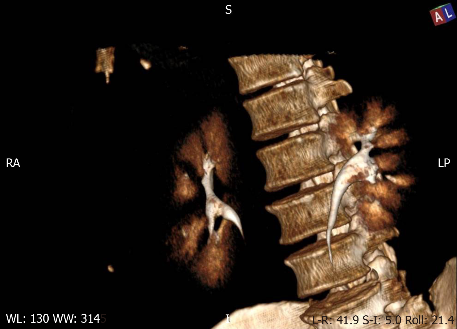
Figure 3 Three-dimensional volume rendering technique of the contrast-enhanced excretory phase.
The image shows distortion of the right kidney major calyces.
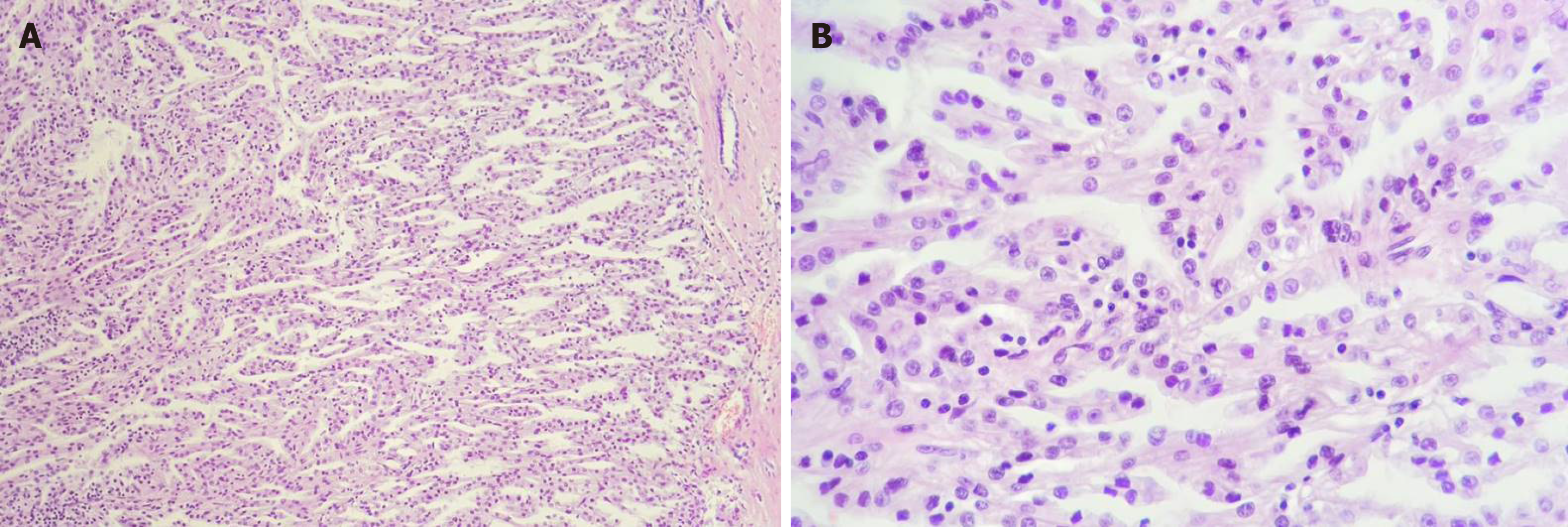
Figure 4 Histological features of the collecting duct carcinoma.
A: Papillary architecture (hematoxylin and eosin, magnification 10 ×); B: Fibrovascular cores lined by one layer of tumor cells, with Fuhrman nuclear grade 2 (hematoxylin and eosin, magnification 20 ×).
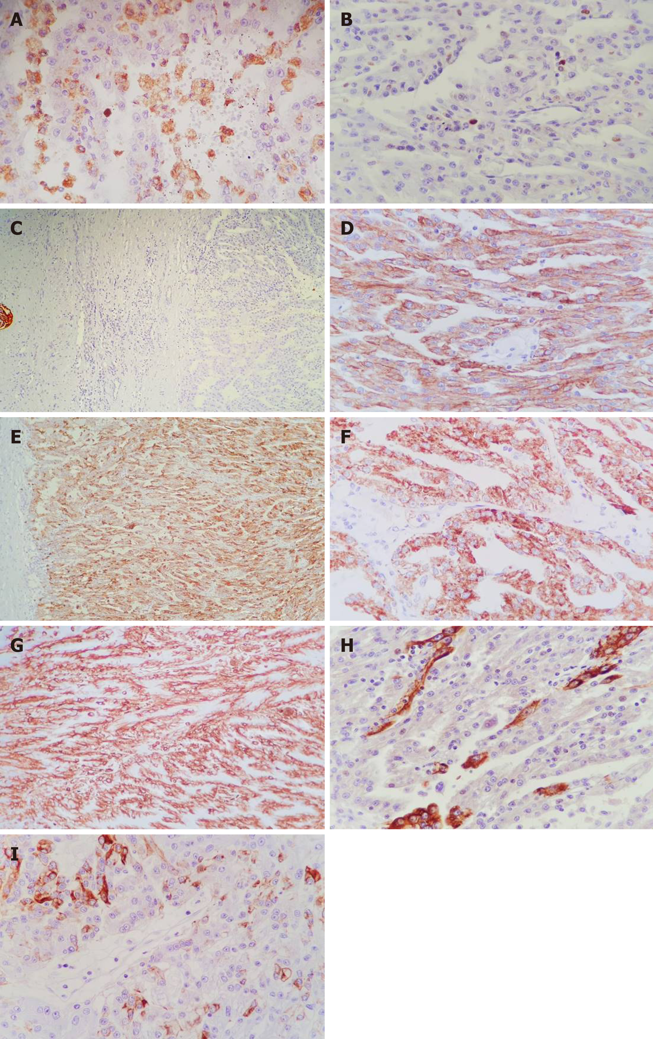
Figure 5 Immunohistochemical profile of the collecting duct carcinoma.
A: Tumor papillae containing CD68-positive foamy macrophages (magnification 20 ×); B: Proliferation index Ki67 < 5% (magnification 20 ×); C: CD10 – no expression on the tumor cells, with positive internal control on the left (magnification 4 ×); D: CK AE1/AE3 – diffuse positivity (magnification 20 ×); E: CD15 – diffuse positivity (magnification 10 ×); F: AMACR – diffuse positivity (magnification 10 ×); G: Vimentin – diffuse positivity (magnification 10 ×); H: CK19 – focal expression (magnification 20 ×); I: CK7 – focal expression (magnification 20 ×).
- Citation: Fulop ZZ, Gurzu S, Jung I, Simu P, Banias L, Fulop E, Dragus E, Bara TJ. Cystic low-grade collecting duct renal carcinoma with liver compression — A challenging diagnosis and therapy: A case report. World J Gastrointest Surg 2020; 12(6): 298-306
- URL: https://www.wjgnet.com/1948-9366/full/v12/i6/298.htm
- DOI: https://dx.doi.org/10.4240/wjgs.v12.i6.298


