Copyright
©The Author(s) 2020.
World J Gastrointest Surg. Apr 27, 2020; 12(4): 178-189
Published online Apr 27, 2020. doi: 10.4240/wjgs.v12.i4.178
Published online Apr 27, 2020. doi: 10.4240/wjgs.v12.i4.178
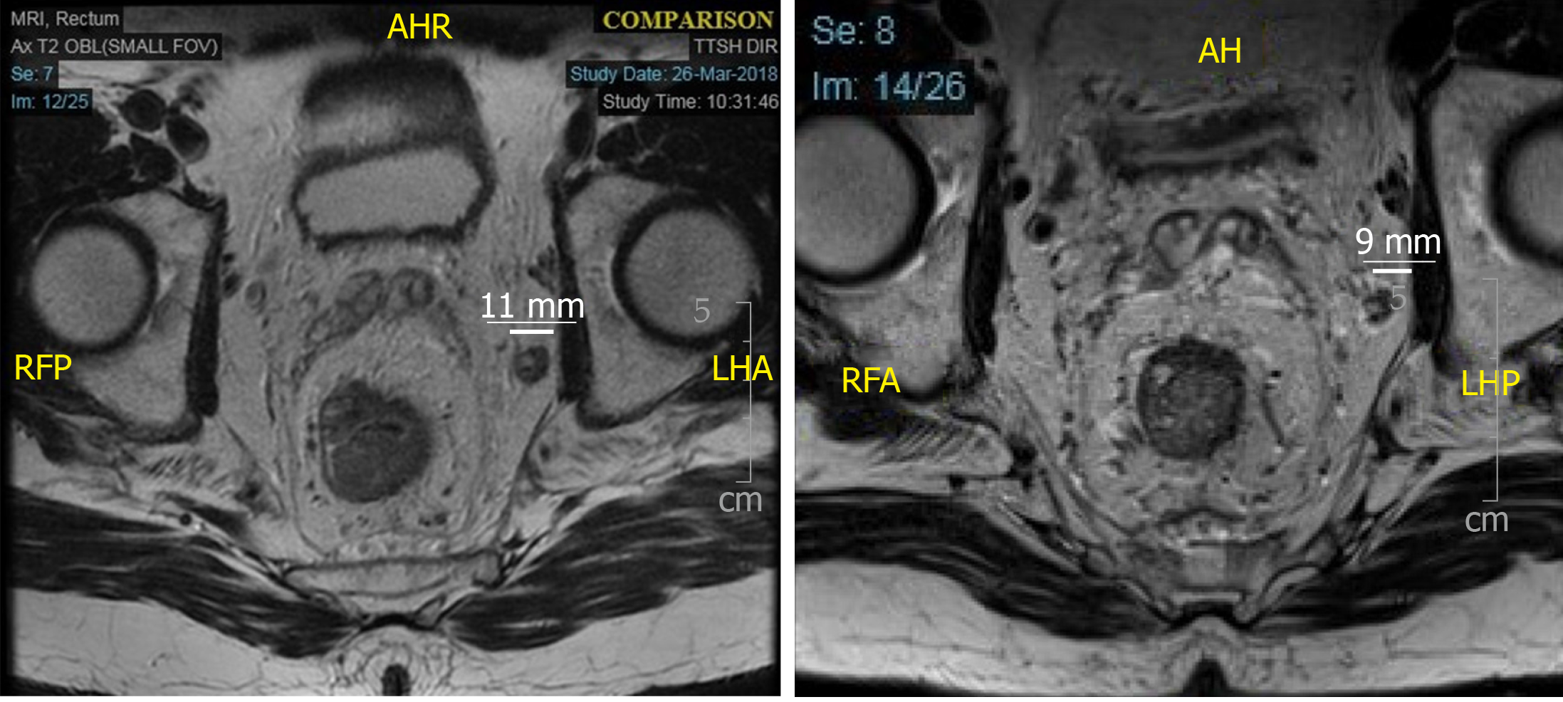
Figure 1 Magnetic resonance imaging rectum showing an enlarged, metastatic, left internal iliac node before (left, 11 mm) and after (right, 9 mm) neoadjuvant chemoradiation.
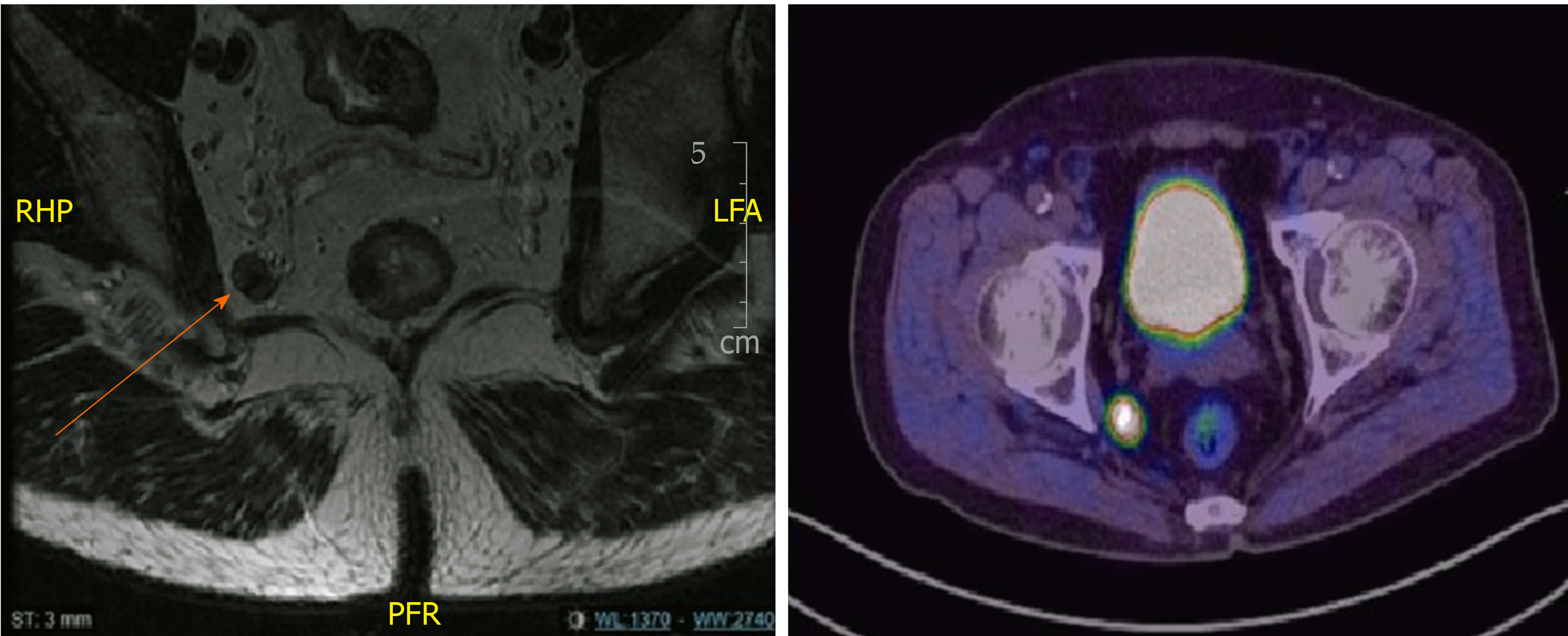
Figure 2 Metastatic lateral pelvic lymph node seen on magnetic resonance imaging rectum and positron emission tomography scan.
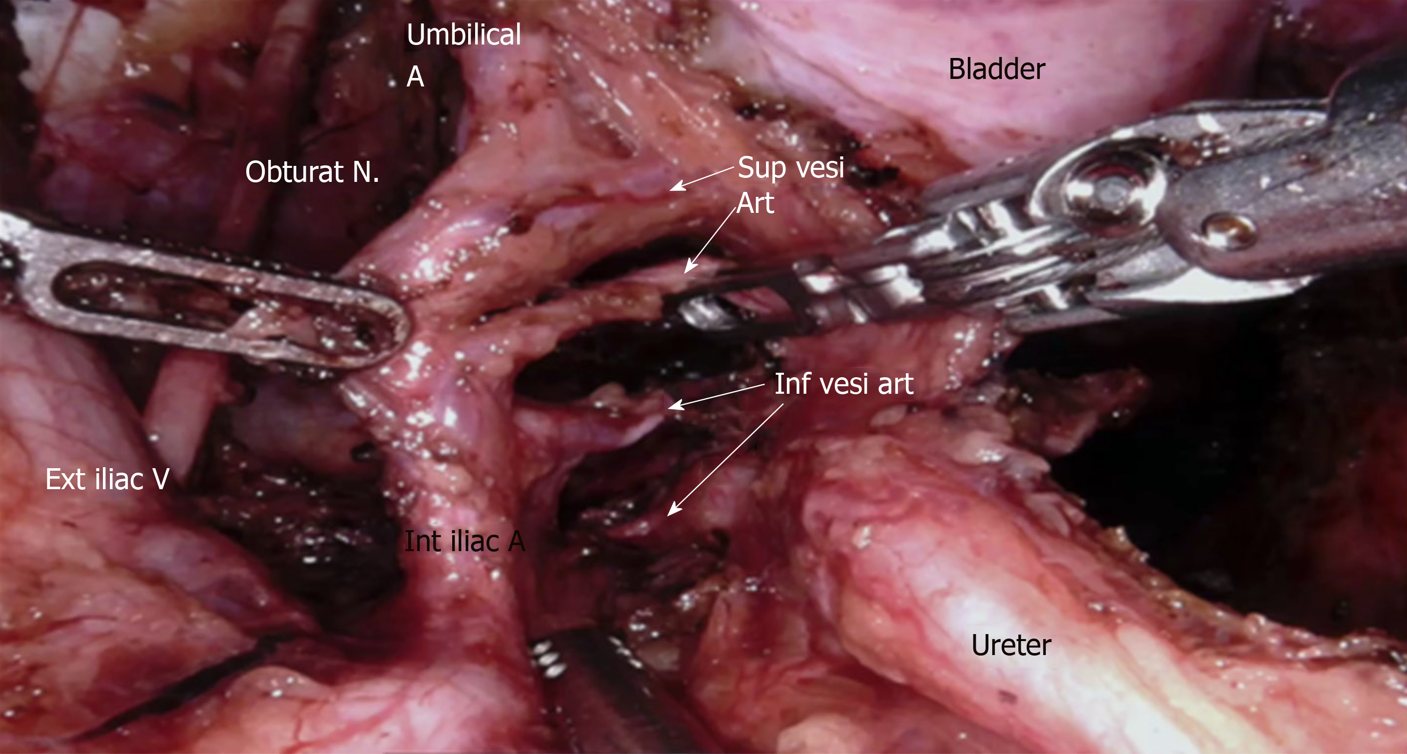
Figure 3 Branches of the internal iliac artery during lateral pelvic lymph node dissection.
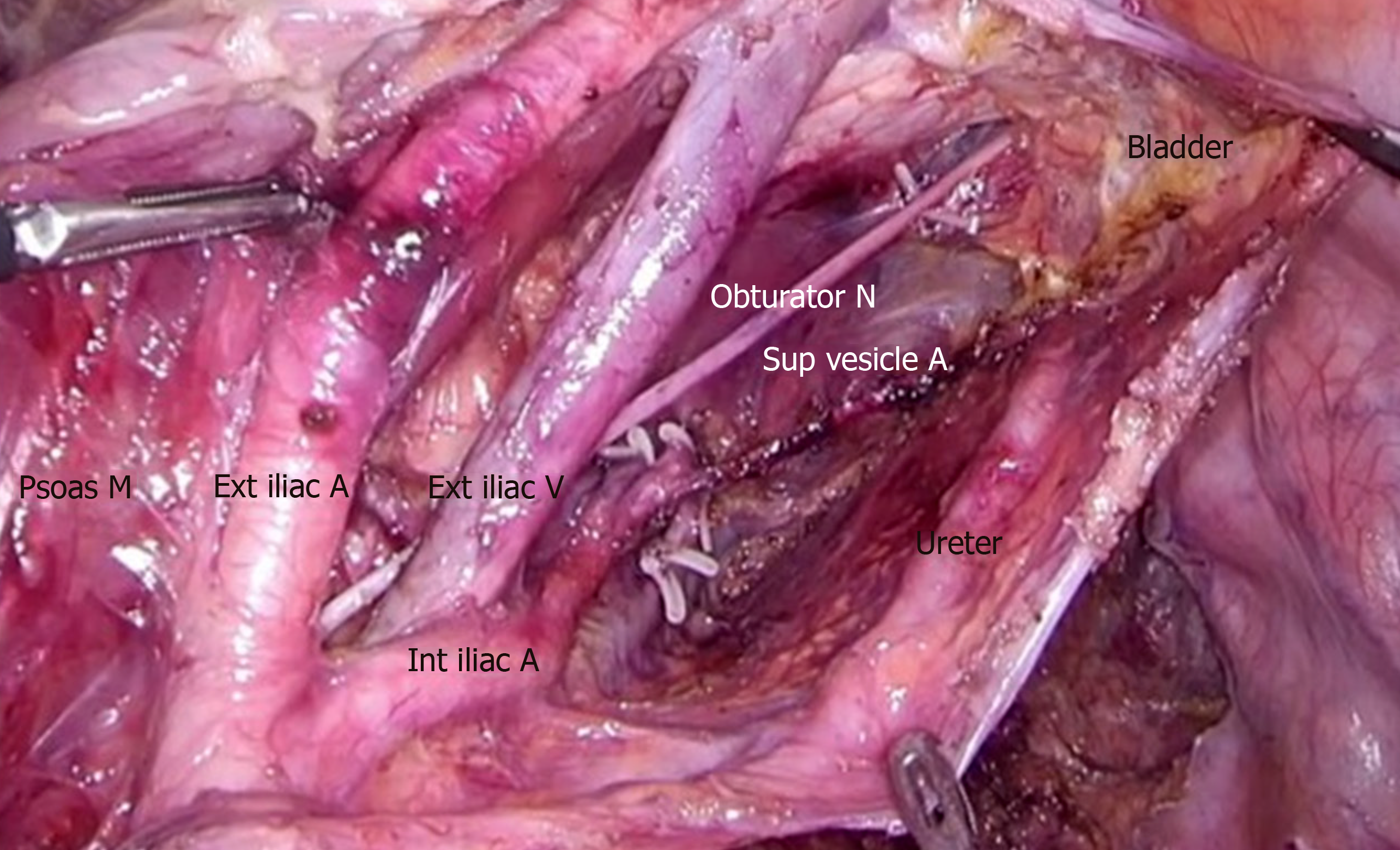
Figure 4 After completion of lateral pelvic lymph node dissection with clearance of external iliac, internal iliac and obturator compartments.
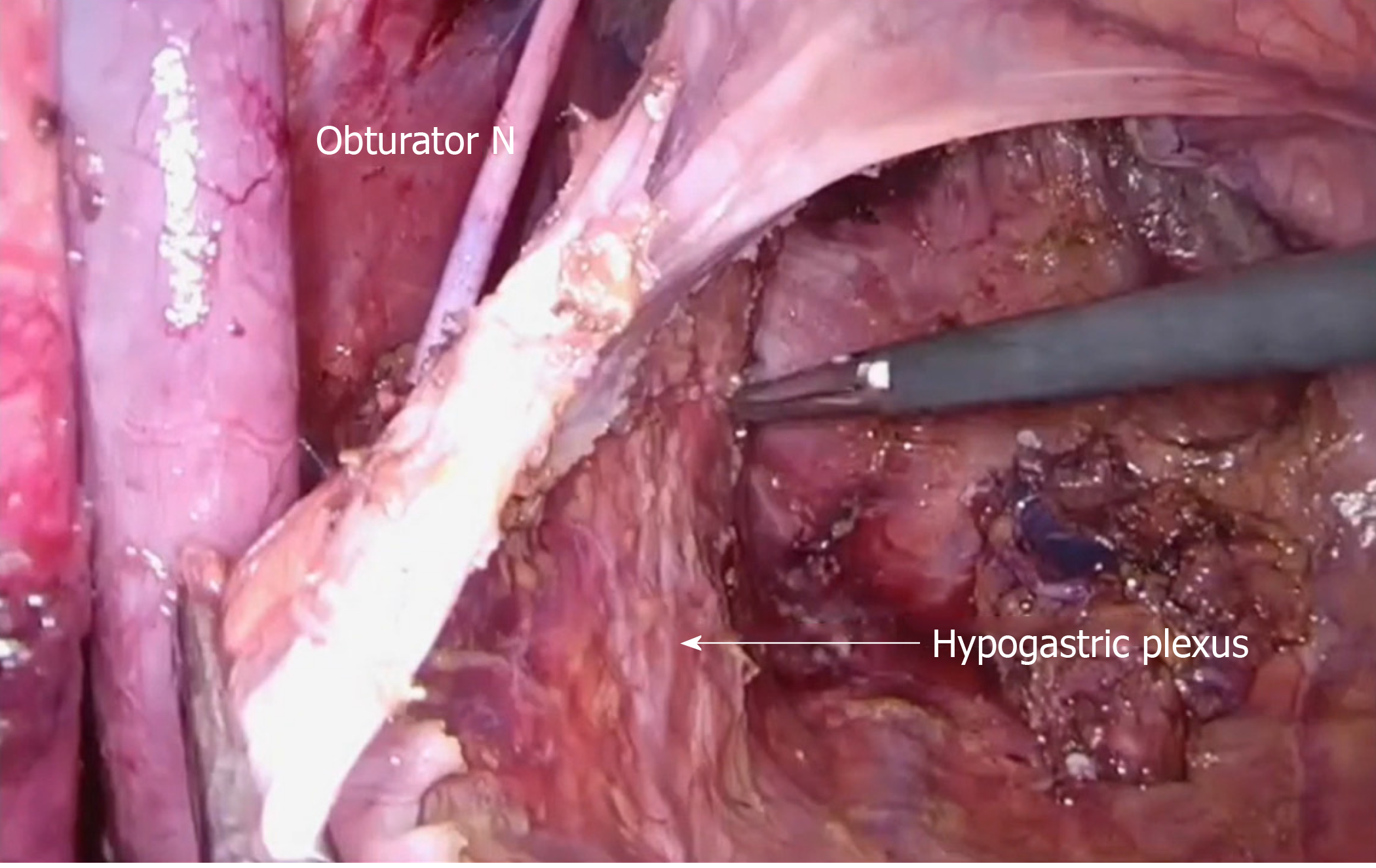
Figure 5 Preservation of ureterohypogastric fascia after lateral pelvic lymph node dissection.
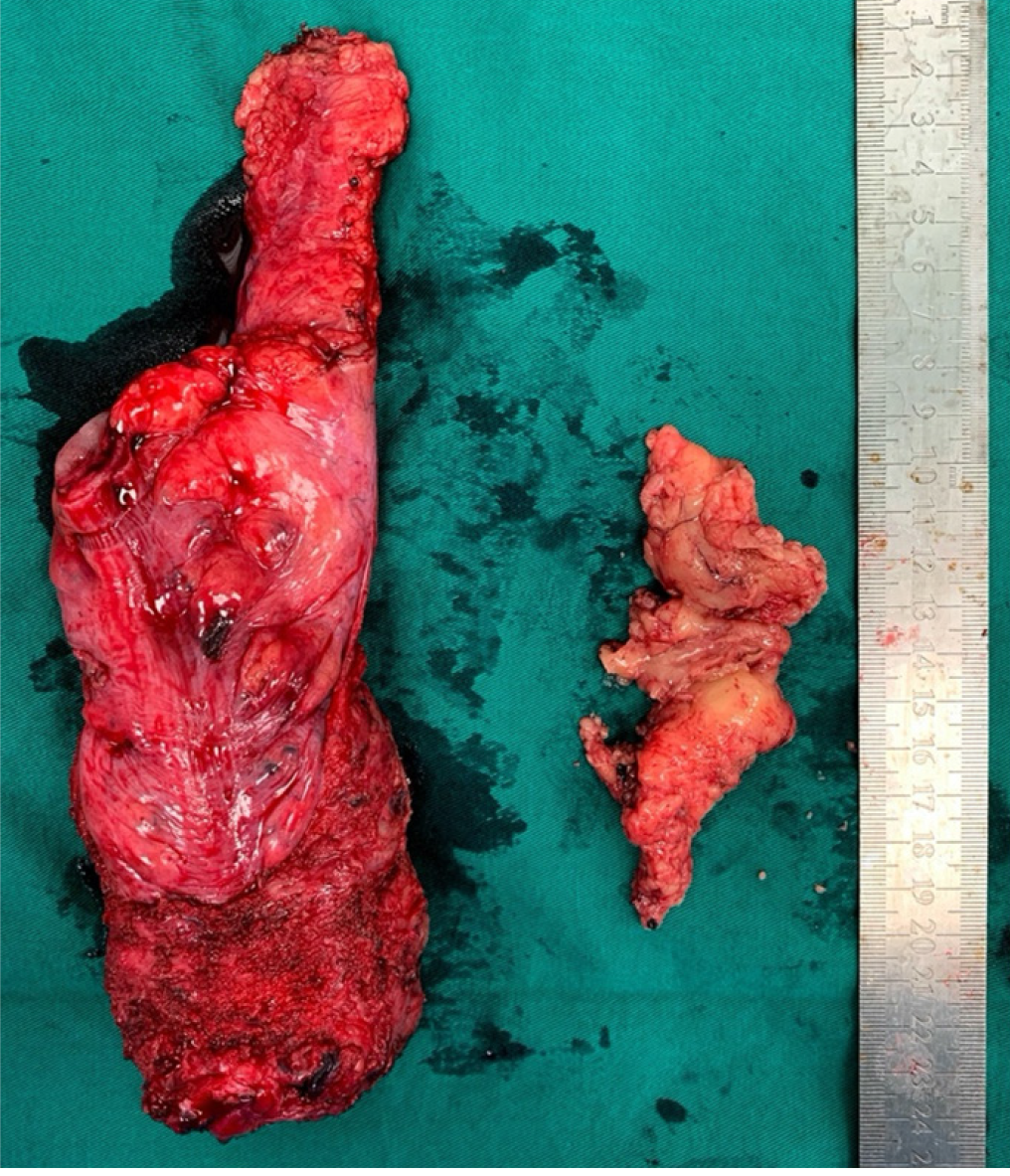
Figure 6 En bloc lymphofatty tissue from lateral pelvic lymph node dissection.
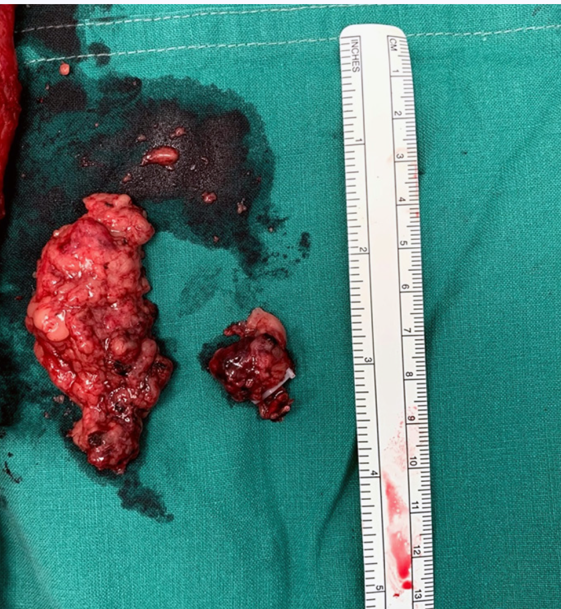
Figure 7 En bloc lymphofatty tissue and metastatic lateral pelvic lymph node.
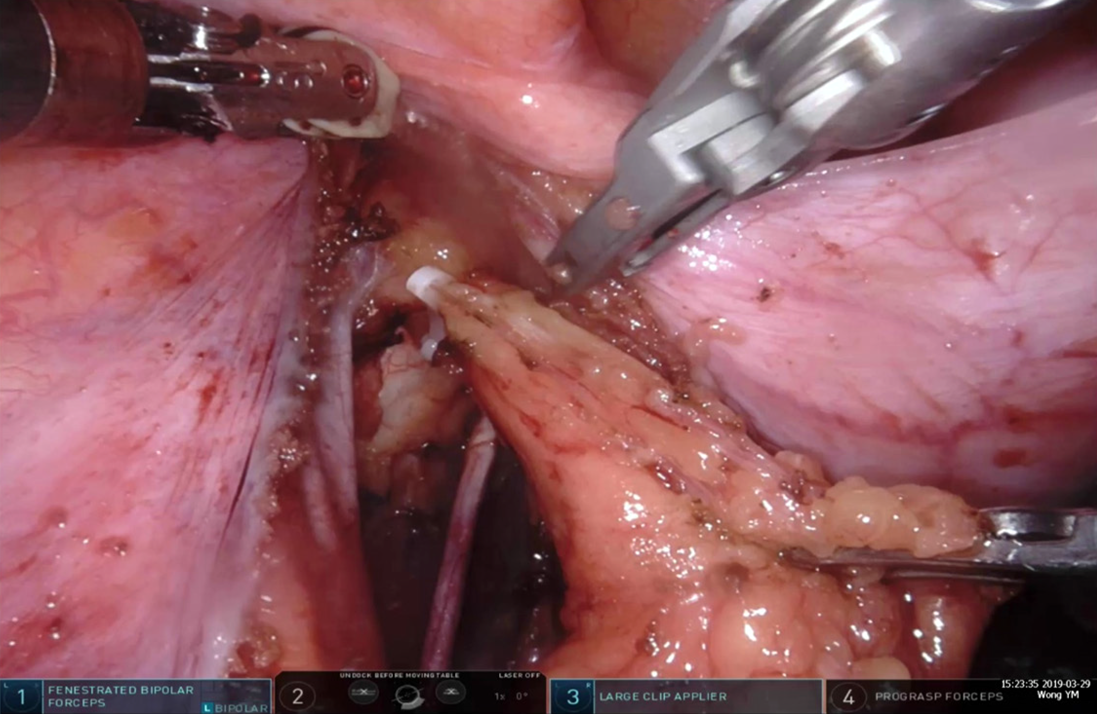
Figure 8 Clipping of a lymphatic channel.
- Citation: Wong KY, Tan AM. Short term outcomes of minimally invasive selective lateral pelvic lymph node dissection for low rectal cancer. World J Gastrointest Surg 2020; 12(4): 178-189
- URL: https://www.wjgnet.com/1948-9366/full/v12/i4/178.htm
- DOI: https://dx.doi.org/10.4240/wjgs.v12.i4.178


