Copyright
©The Author(s) 2020.
World J Gastrointest Surg. Feb 27, 2020; 12(2): 77-84
Published online Feb 27, 2020. doi: 10.4240/wjgs.v12.i2.77
Published online Feb 27, 2020. doi: 10.4240/wjgs.v12.i2.77
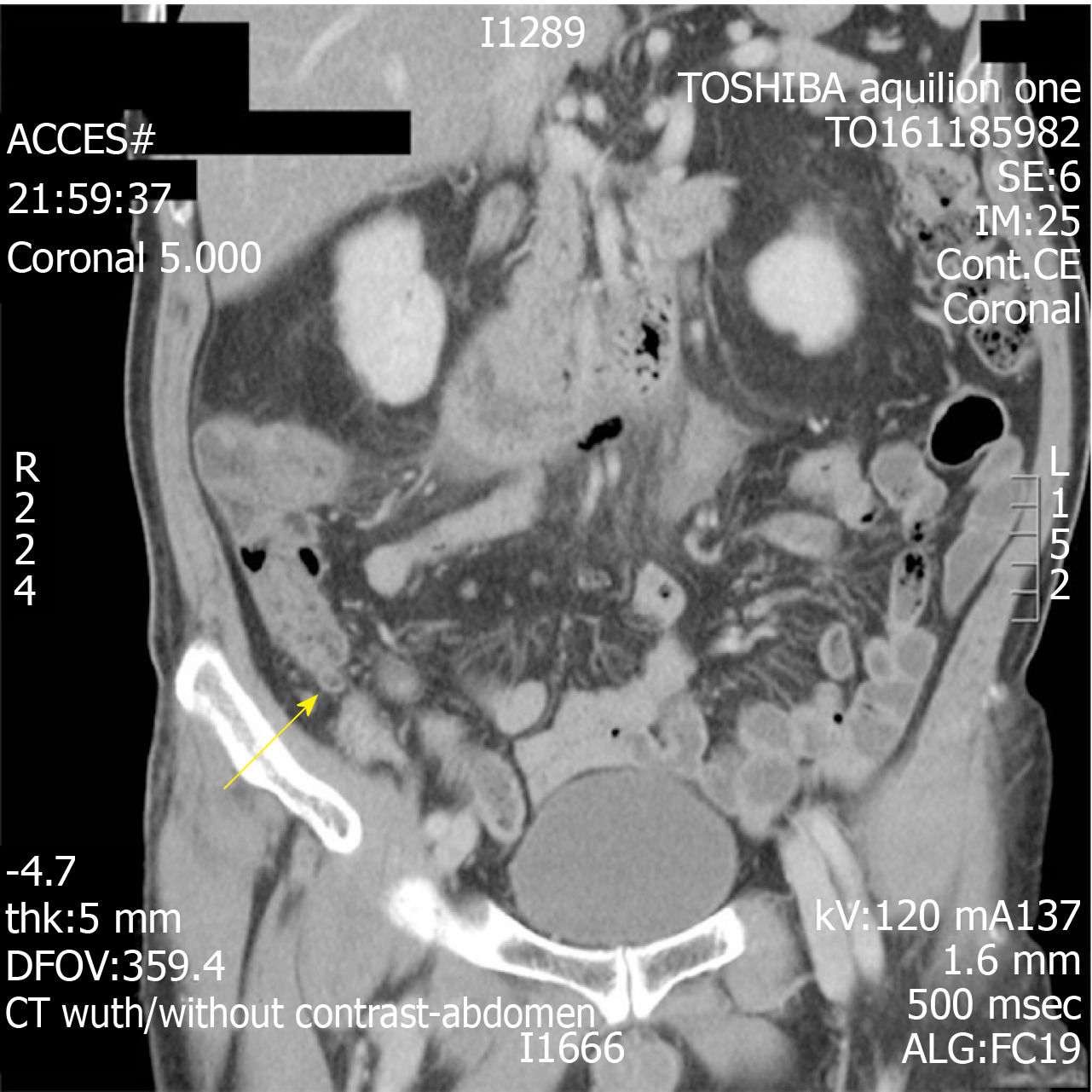
Figure 1 Computed tomography abdomen and pelvic with contrast showing distended tubular structure (demonstrated by the arrow) that was presumed to be the appendix.
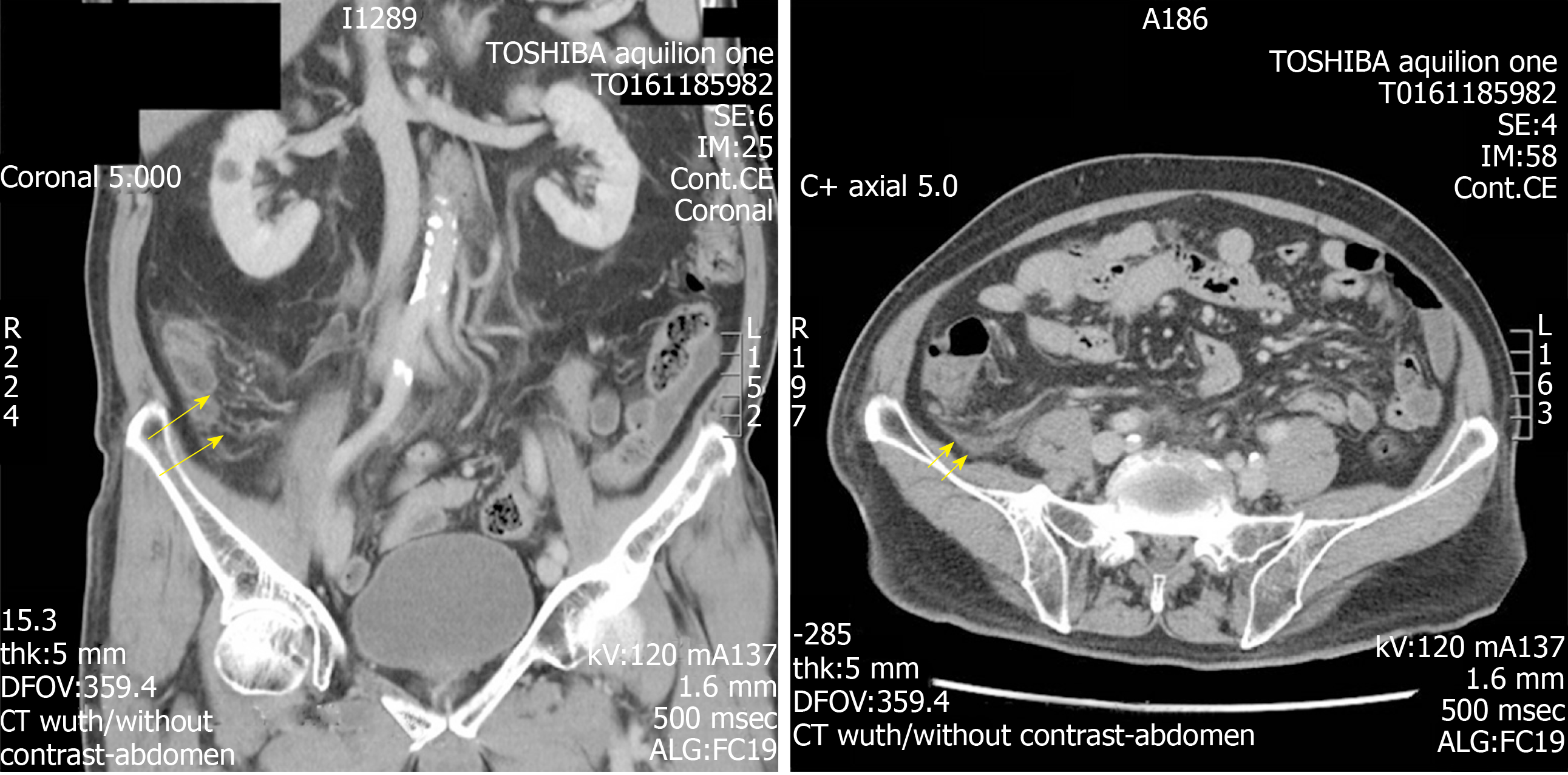
Figure 2 Axial and coronal views of prominent fat stranding in the right lower quadrant (arrows).
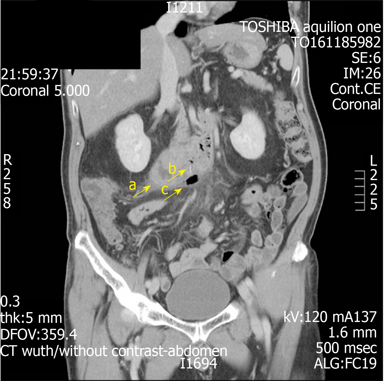
Figure 3 Computed tomography scan showing identified fishbone in the third segment of the duodenum measuring 1.
4 cm long with associated air within the retroperitoneal space (arrow a). Local abscess was noted (arrow b). Area of gas out with the GI tract (arrow c).
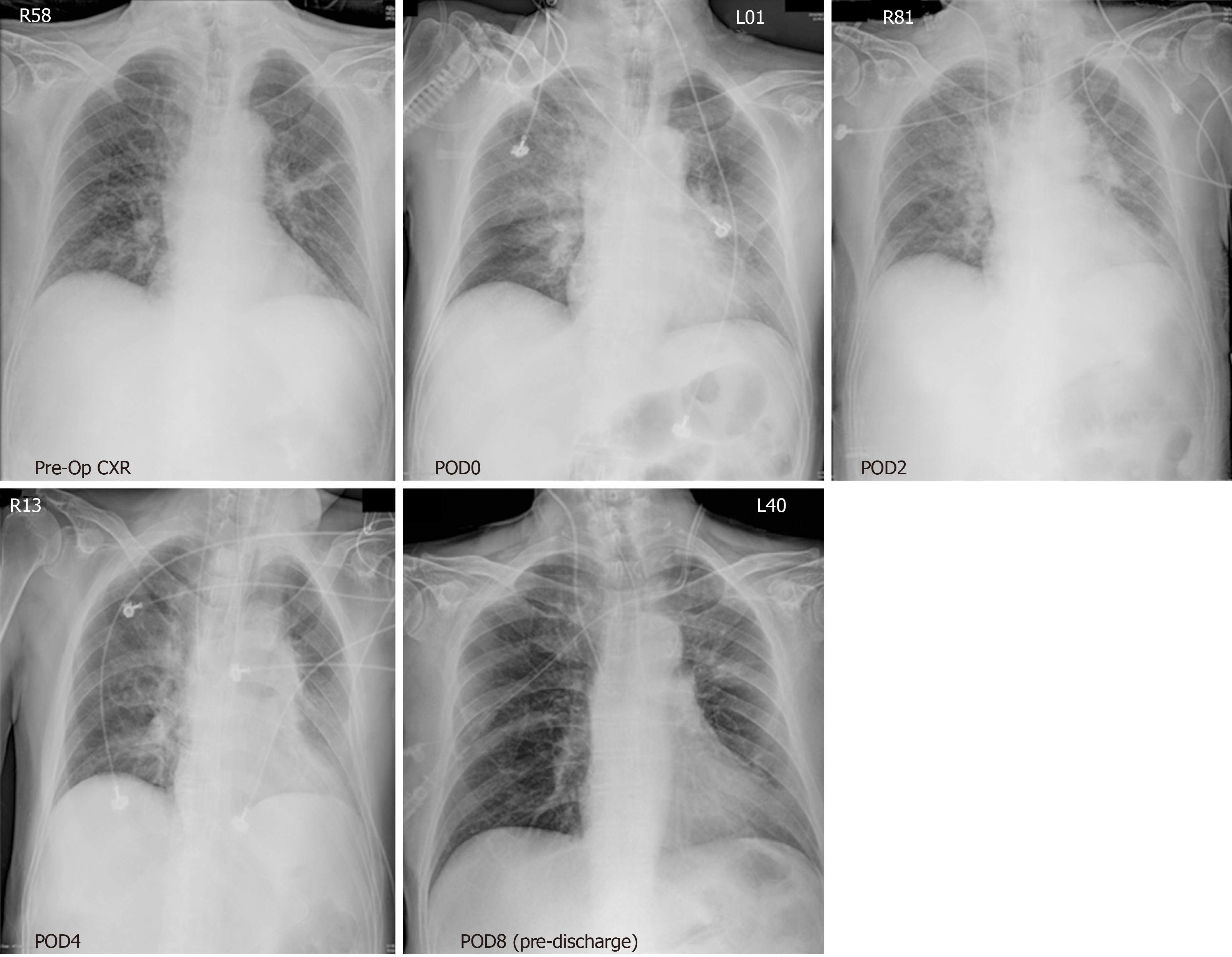
Figure 4 Serial chest radiographs showed acute consolidation and rapid resolution in his right lobe during the span of his inpatient stay.
View from top left to bottom right. POD: Post-operative day.
- Citation: Lim D, Ho CM. Appendicitis-mimicking presentation in fishbone induced microperforation of the distal duodenum: A case report. World J Gastrointest Surg 2020; 12(2): 77-84
- URL: https://www.wjgnet.com/1948-9366/full/v12/i2/77.htm
- DOI: https://dx.doi.org/10.4240/wjgs.v12.i2.77


