Copyright
©The Author(s) 2020.
World J Gastrointest Surg. Oct 27, 2020; 12(10): 435-441
Published online Oct 27, 2020. doi: 10.4240/wjgs.v12.i10.435
Published online Oct 27, 2020. doi: 10.4240/wjgs.v12.i10.435
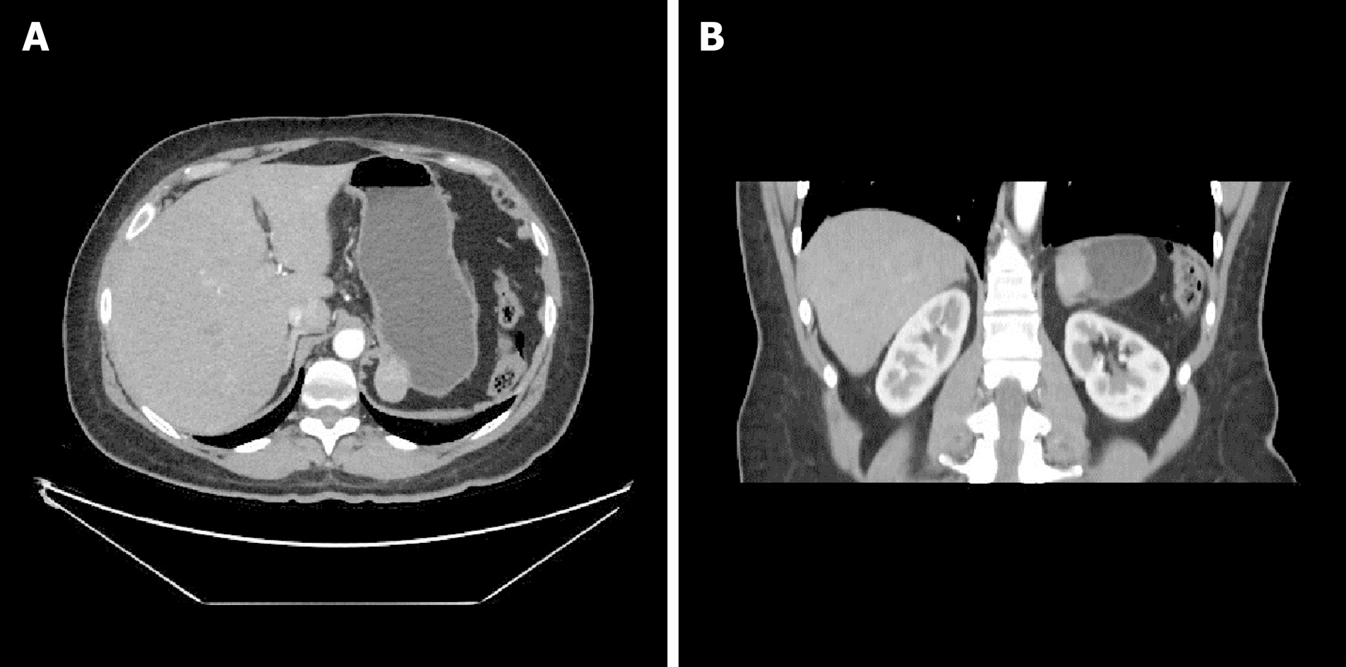
Figure 1 Preoperative abdominal computed tomography scan with intravenous contrast administration: A: Transverse; B: Coronal.
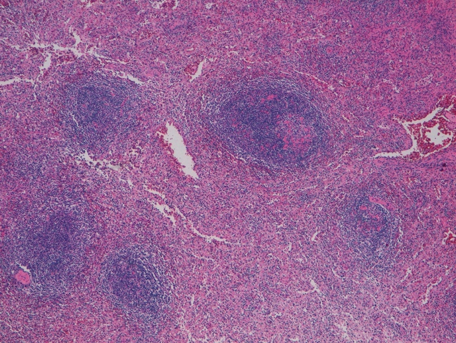
Figure 2 Lymphoid tissue found in the gastric nodule (hematoxylin and eosin staining, ×4).
- Citation: Isopi C, Vitali G, Pieri F, Solaini L, Ercolani G. Gastric splenosis mimicking a gastrointestinal stromal tumor: A case report. World J Gastrointest Surg 2020; 12(10): 435-441
- URL: https://www.wjgnet.com/1948-9366/full/v12/i10/435.htm
- DOI: https://dx.doi.org/10.4240/wjgs.v12.i10.435


