Copyright
©The Author(s) 2019.
World J Gastrointest Surg. Sep 27, 2019; 11(9): 358-364
Published online Sep 27, 2019. doi: 10.4240/wjgs.v11.i9.358
Published online Sep 27, 2019. doi: 10.4240/wjgs.v11.i9.358
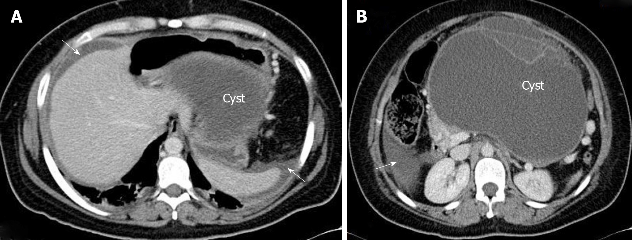
Figure 1 A 37-year-old woman operated on for a ruptured giant mucinous cystadenoma of the pancreas.
A: Computed tomography image showing free mucinous ascites (arrows) and the dome of the pancreatic cystic lesion (mucinous cystadenoma with focal adenocarcinoma) in the retrogastric area; B: Peritoneal fluid in the right paracolic gutter (arrow) and corporal cystic lesion with septa.
Figure 2 Thoracic computed tomography images showing bilateral nodular metastases (arrows) from a low-grade appendiceal mucinous neoplasm.
Figure 3 Two forms of omental cake in two patients with peritoneal dissemination of low-grade appendiceal mucinous neoplasm.
Note the sparing of the small bowel in the right picture.
- Citation: Morera-Ocon FJ, Navarro-Campoy C. History of pseudomyxoma peritonei from its origin to the first decades of the twenty-first century. World J Gastrointest Surg 2019; 11(9): 358-364
- URL: https://www.wjgnet.com/1948-9366/full/v11/i9/358.htm
- DOI: https://dx.doi.org/10.4240/wjgs.v11.i9.358


