Copyright
©The Author(s) 2019.
World J Gastrointest Surg. Aug 27, 2019; 11(8): 342-347
Published online Aug 27, 2019. doi: 10.4240/wjgs.v11.i8.342
Published online Aug 27, 2019. doi: 10.4240/wjgs.v11.i8.342
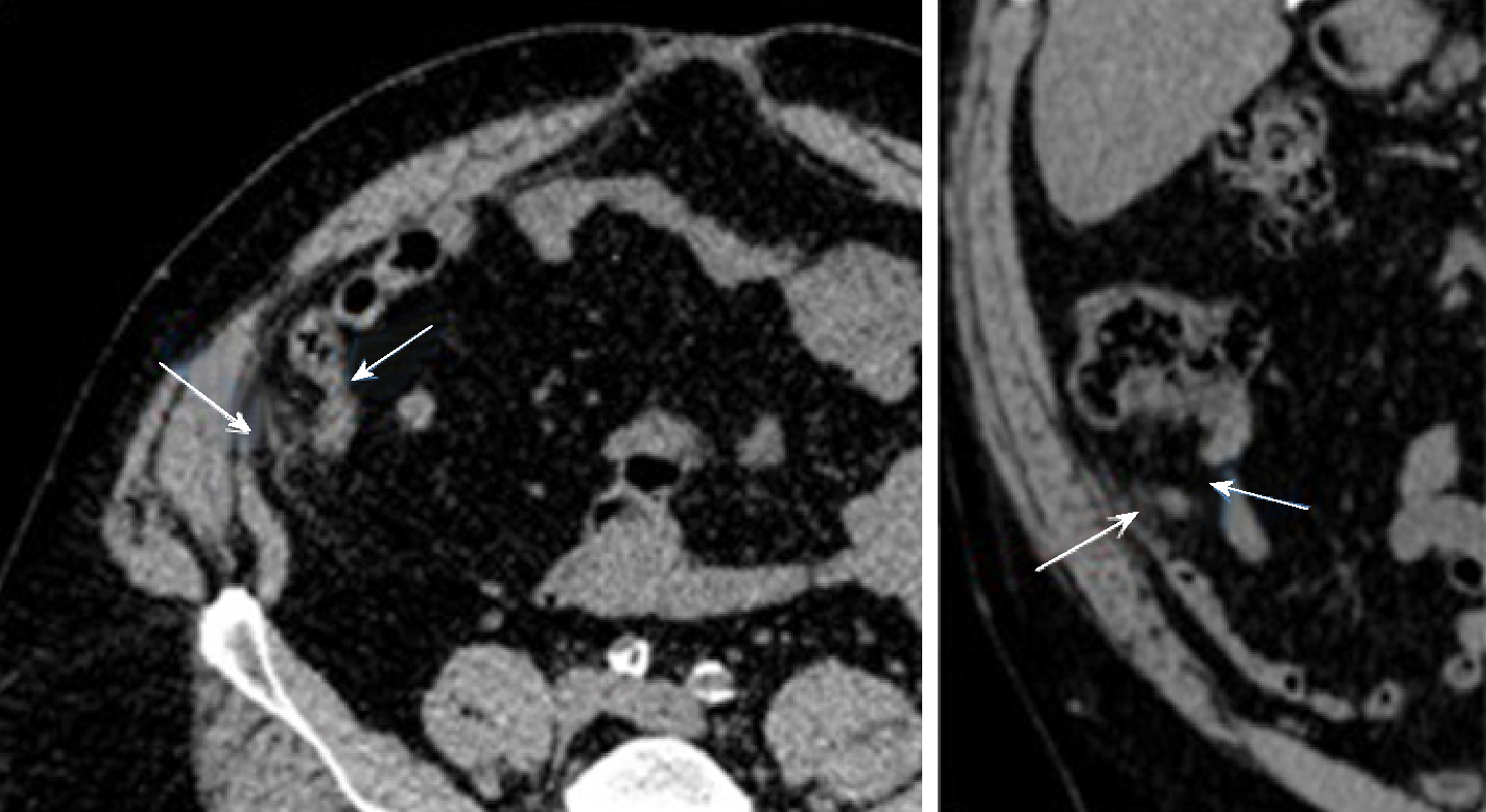
Figure 1 Abdominal computed tomography scan.
A 1.0 cm × 1.8 cm focus of oval inflammatory changes surrounding central fat density visualized adjacent to the tip of the appendix and inferior aspect of the cecum noted. This is likely due to epiploic appendagitis. Possibility of very early acute distal tip appendicitis cannot be entirely excluded but felt to be less likely (Short arrow: Appendix; Long arrow: Epiploic appendagitis).
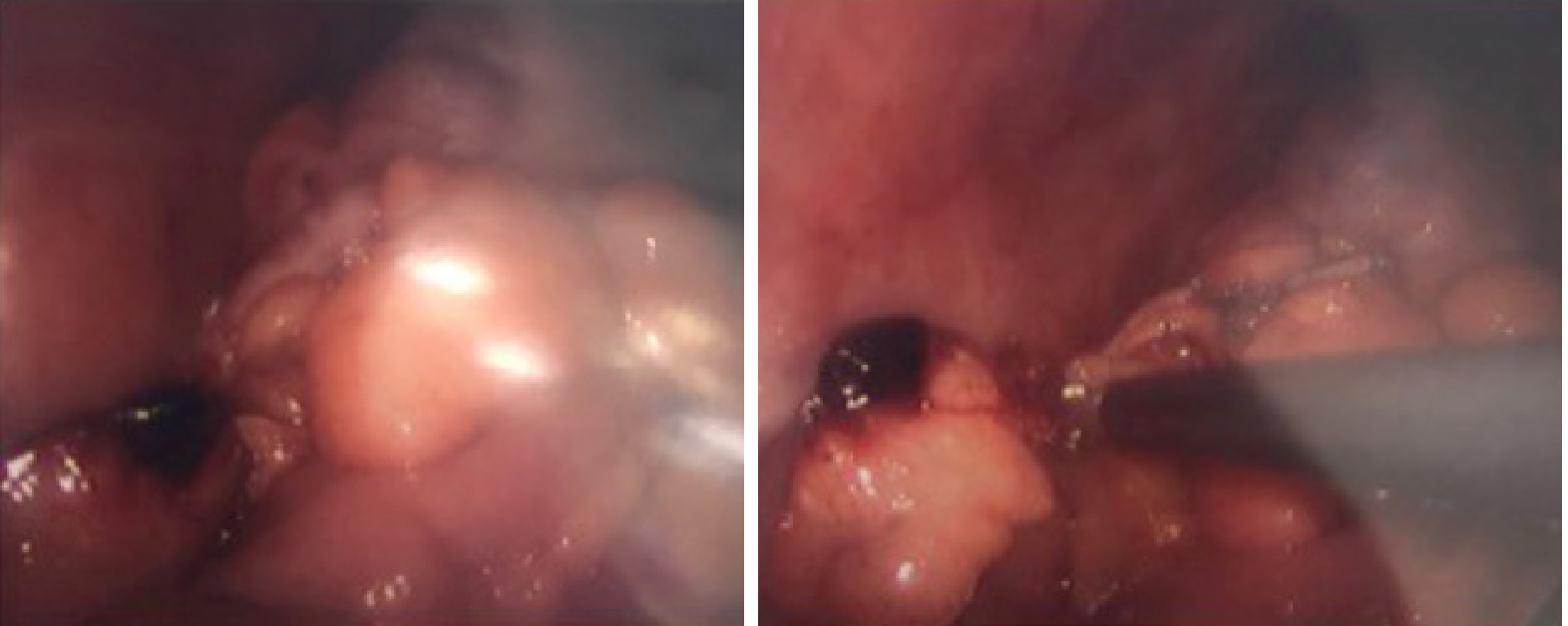
Figure 2 Infarcted appendiceal epiploic appendage at the tip of the appendix (Intraoperative).
Figure 3 The Congested and hemorrhagic appendage.
A: The congested and hemorrhagic appendage measures 6.3 cm × 1.6 cm × 1 cm; B: Serosal surface with fibrin and few acute inflammatory cells. Muscular layer with no inflammatory cells. High power.
- Citation: Huang K, Waheed A, Juan W, Misra S, Alpendre C, Jones S. Acute epiploic appendagitis at the tip of the appendix mimicking acute appendicitis: A rare case report with literature review. World J Gastrointest Surg 2019; 11(8): 342-347
- URL: https://www.wjgnet.com/1948-9366/full/v11/i8/342.htm
- DOI: https://dx.doi.org/10.4240/wjgs.v11.i8.342


