Copyright
©The Author(s) 2019.
World J Gastrointest Surg. Mar 27, 2019; 11(3): 191-197
Published online Mar 27, 2019. doi: 10.4240/wjgs.v11.i3.191
Published online Mar 27, 2019. doi: 10.4240/wjgs.v11.i3.191
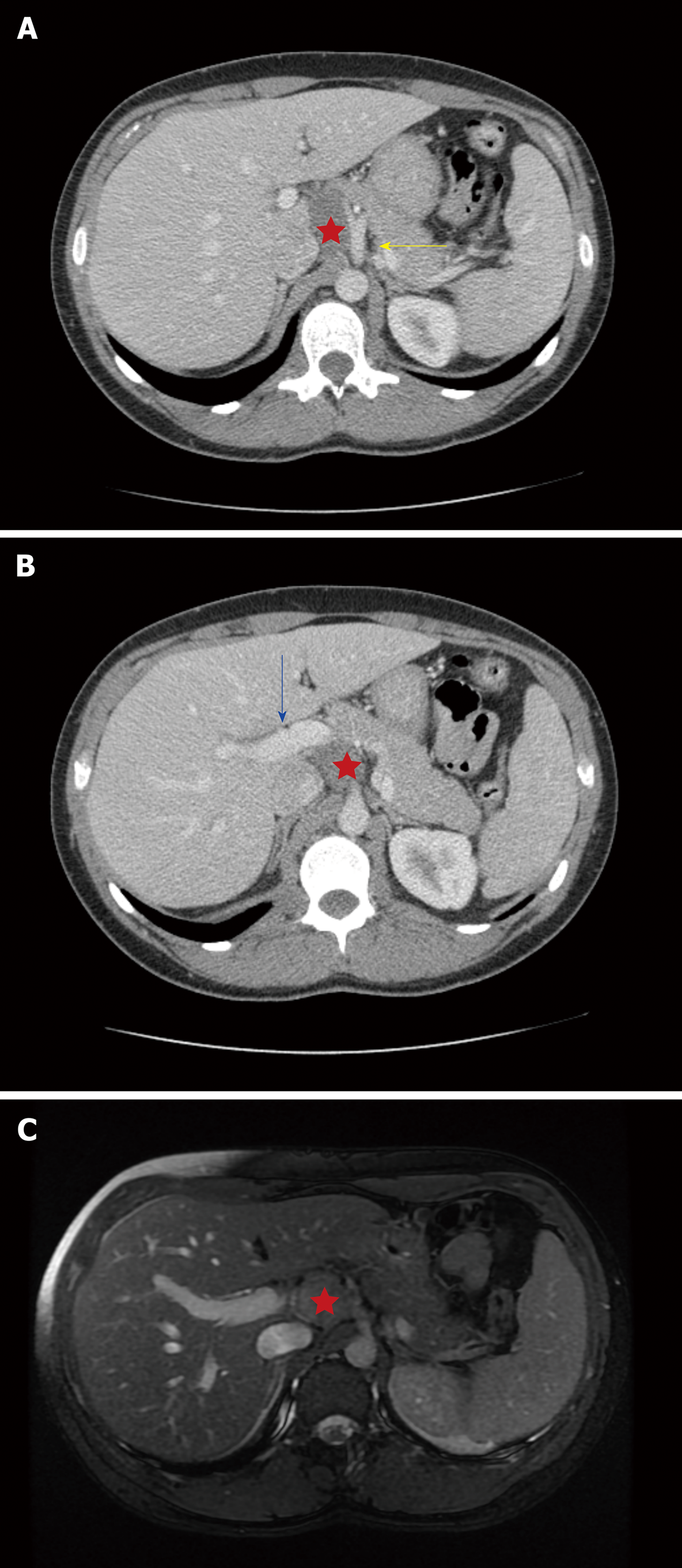
Figure 1 Axial computed tomography scan and magnetic resonance imaging.
A: Axial computed tomography (CT) scan: This axial CT image demonstrates the ganglioneuroma (red star) abutting the celiac axis (yellow arrow) and the caudate lobe of the liver; B: Axial CT scan: A more inferior axial CT cut demonstrates the inferomedial extension of the ganglioneuroma (red star) adjacent to the portal vein (blue arrow); C: Axial magnetic resonance imaging (MRI): This MRI image demonstrates the close proximity of the ganglioneuroma (red star) to the vital structures. However, there was no vascular encasement or invasion and fat planes were preserved.
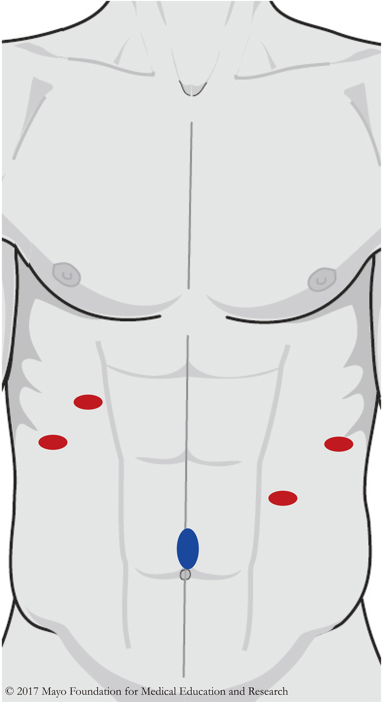
Figure 2 Port placement.
This diagram shows the port placement during the operation. The Hasson trocar (blue oval) was first placed in a supraumbilical position. Four additional 5 mm trocars (red ovals) were placed under direct vision and the right lateral subcostal port was used for the liver retractor. Image was created with permission from Mayo Foundation for Medical Education and Research.
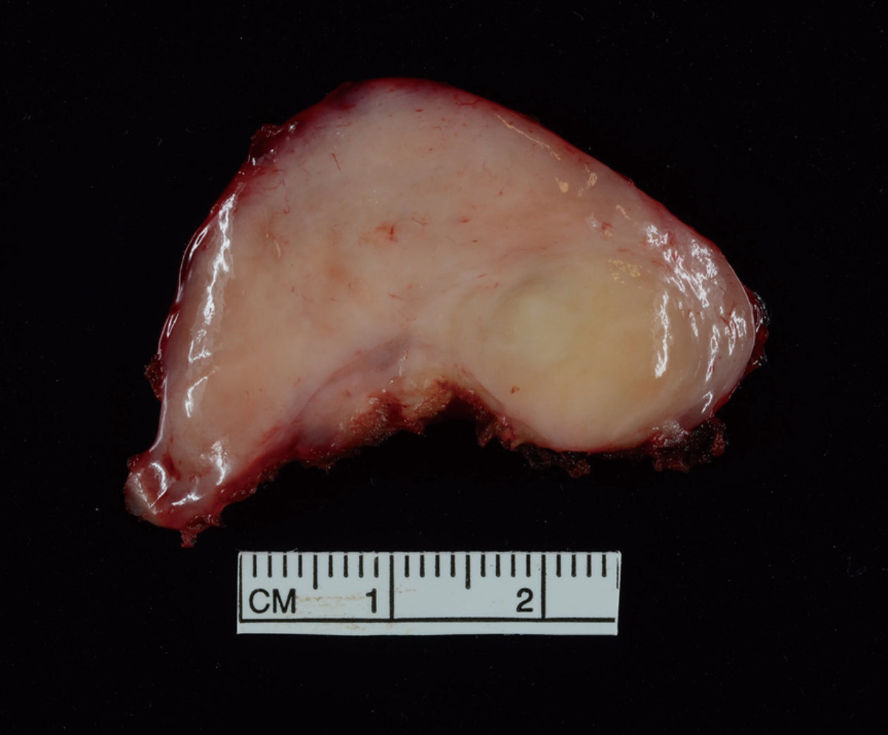
Figure 3 Pathology specimen.
The pathology specimen shows a lobulated, well-circumscribed ganglioneuroma (43 mm in greatest dimension).
- Citation: Hemmati P, Ghanem O, Bingener J. Laparoscopic celiac plexus ganglioneuroma resection: A video case report. World J Gastrointest Surg 2019; 11(3): 191-197
- URL: https://www.wjgnet.com/1948-9366/full/v11/i3/191.htm
- DOI: https://dx.doi.org/10.4240/wjgs.v11.i3.191


