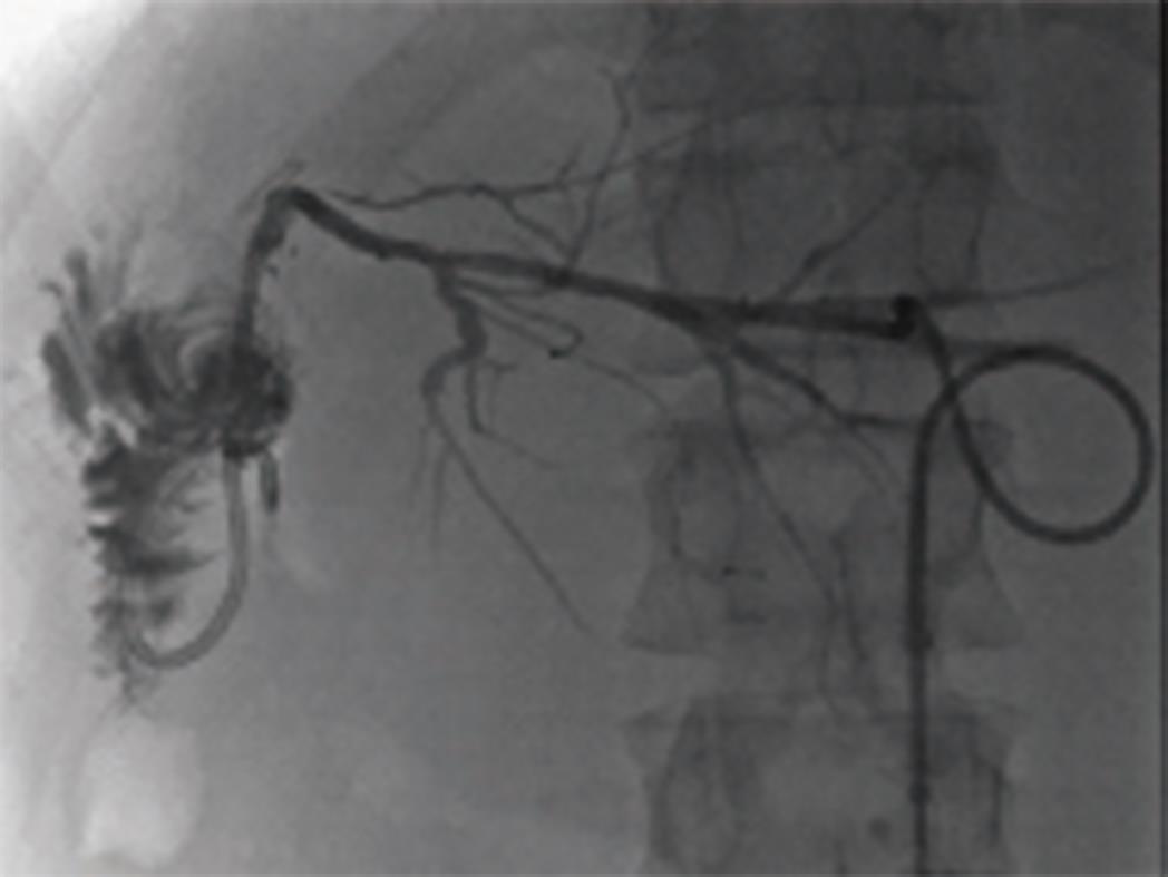
Copyright
©The Author(s) 2018.
World J Gastrointest Surg. Jan 27, 2018; 10(1): 1-5
Published online Jan 27, 2018. doi: 10.4240/wjgs.v10.i1.1
Published online Jan 27, 2018. doi: 10.4240/wjgs.v10.i1.1
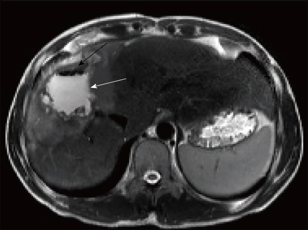
Figure 1 T2 axial dynamic liver magnetic resonance imaging.
A mass lesion with heterogenous signal density that completely occupies segment 4 and right hepatic lobe and contains air densities (black arrow) and infected collection (white arrow).
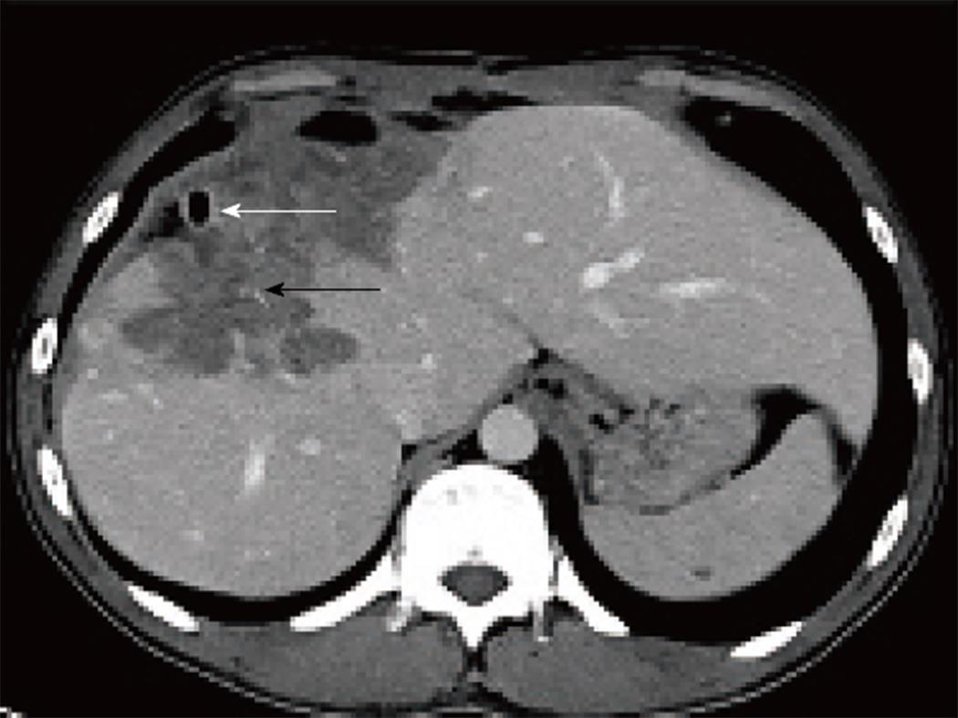
Figure 2 Preoperative axial venous phase multidetector computerized tomography image.
A percutaneous drainage catheter to drain the infected content of the lesion is seen (white arrow). Furthermore, it observed that the cystic lesion causes a retraction in the hepatic capsule and shows invasion to surrounding tissue with millimetric amorphous calcification foci (black arrow).
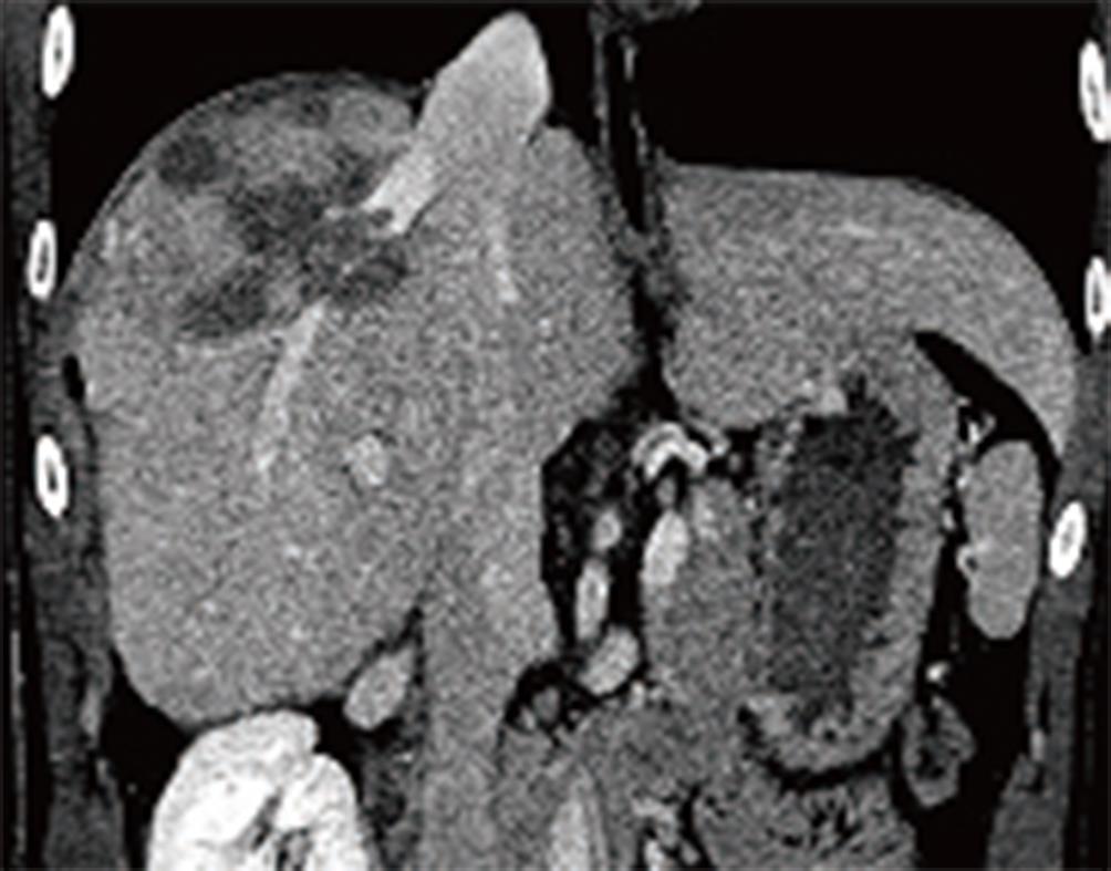
Figure 3 Preoperative reconstructed coronal multidetector computerized tomography images.
The lesion is invading the right hepatic vein but did not extend to the retrohepatic inferior vena cava.
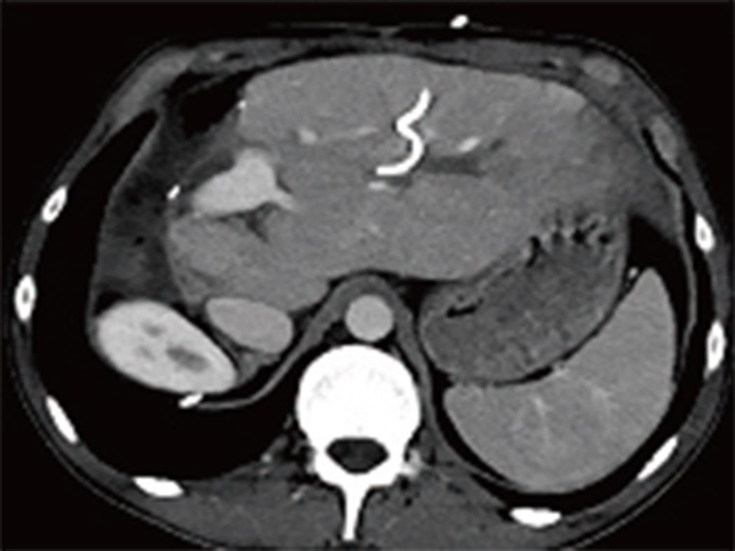
Figure 4 Postoperative axial phase multidetector computerized tomography image.
Extended right hepatectomy involving the right lobe and segment IV is performed remaining the segment II and III. Segment III bile duct is dilated (white arrow) and an external biliary drainage catheter is seen inside the bile duct (black arrow).
Figure 5 Intraoperative cholangiography through the external biliary drainage catheter following hepaticojejunostomy is seen.
The images following contrast injection demonstrates patent bile ducts and no leakage from the anastomosis site.
- Citation: Akbulut S, Cicek E, Kolu M, Sahin TT, Yilmaz S. Associating liver partition and portal vein ligation for staged hepatectomy for extensive alveolar echinococcosis: First case report in the literature. World J Gastrointest Surg 2018; 10(1): 1-5
- URL: https://www.wjgnet.com/1948-9366/full/v10/i1/1.htm
- DOI: https://dx.doi.org/10.4240/wjgs.v10.i1.1


